
Page 229 - Atlas of Small Animal CT and MRI
P. 229
Infectious Inflammatory Disorders 219
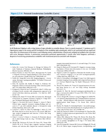
Figure 2.7.14 Protozoal Granulomatous Cerebellitis (Canine) MR
(a) T2, TP (b) T1, TP (c) T1+C, TP
6y M Rhodesian Ridgeback with a 4‐day history of signs referable to cerebellar disease. There is a poorly margined, T1 isointense and T2
hyperintense region in the vermis and left hemisphere of the cerebellum (a,b: arrowheads), which is not associated with any significant
mass effect. No enhancement of the lesion is seen following contrast administration. A fluorescent antibody test yielded a titer consistent
with active Neospora infection. A postmortem examination performed approximately 3 months after initial diagnosis confirmed a
diagnosis of necrotizing granulomatous cerebellitis with intralesional protozoa consistent with Neospora.
References otogenic intracranial infection in 11 cats and 4 dogs. J Vet Intern
Med. 2006;20:648–656.
1. Sellon RK. Canine Viral Diseases. In: Ettinger SJ, Feldman EC 11. Klopp LS, Hathcock JT, Sorjonen DC. Magnetic resonance imag-
(eds): Textbook of Veterinary Internal Medicine. Philadelphia: ing features of brain stem abscessation in two cats. Vet Radiol
Elsevier Saunders, 2005;646–652. Ultrasound. 2000;41:300–307.
2. Bathen‐Noethen A, Stein VM, Puff C, Baumgaertner W, Tipold 12. Negrin A, Lamb CR, Cappello R, Cherubini GB. Results of mag-
A. Magnetic resonance imaging findings in acute canine distem- netic resonance imaging in 14 cats with meningoencephalitis.
per virus infection. J Small Anim Pract. 2008;49:460–467. J Feline Med Surg. 2007;9:109–116.
3. Griffin JFt, Young BD, Levine JM. Imaging diagnosis – chronic 13. Seiler G, Cizinauskas S, Scheidegger J, Lang J. Low‐field magnetic
canine distemper meningoencephalitis. Vet Radiol Ultrasound. resonance imaging of a pyocephalus and a suspected brain abscess
2009;50:182–184. in a German Shepherd dog. Vet Radiol Ultrasound. 2001;42:
4. Foley JE, Lapointe JM, Koblik P, Poland A, Pedersen NC. 417–422.
Diagnostic features of clinical neurologic feline infectious perito- 14. Wouters EG, Beukers M, Theyse LF. Surgical treatment of a cere-
nitis. J Vet Intern Med. 1998;12:415–423. bral brain abscess in a cat. Vet Comp Orthop Traumatol.
5. Kitagawa M, Okada M, Sato T, Kanayama K, Sakai T. A feline case 2011;24:72–75.
of isolated fourth ventricle with syringomyelia suspected to be 15. Radaelli ST, Platt SR. Bacterial meningoencephalomyelitis in
related with feline infectious peritonitis. J Vet Med Sci. 2007; dogs: a retrospective study of 23 cases (1990–1999). J Vet Intern
69:759–762. Med. 2002;16:159–163.
6. Mellema LM, Samii VF, Vernau KM, LeCouteur RA. Meningeal 16. Anor S, Sturges BK, Lafranco L, Jang SS, Higgins RJ, Koblik PD,
enhancement on magnetic resonance imaging in 15 dogs and 3 et al. Systemic phaeohyphomycosis (Cladophialophora bantiana)
cats. Vet Radiol Ultrasound. 2002;43:10–15. in a dog – clinical diagnosis with stereotactic computed tomo-
7. Negrin A, Cherubini GB, Lamb C, Benigni L, Adams V, Platt S. graphic‐guided brain biopsy. J Vet Intern Med. 2001;15:
Clinical signs, magnetic resonance imaging findings and out- 257–261.
come in 77 cats with vestibular disease: a retrospective study. 17. Bentley RT, Faissler D, Sutherland‐Smith J. Successful manage-
J Feline Med Surg. 2010;12:291–299. ment of an intracranial phaeohyphomycotic fungal granuloma in
8. Costanzo C, Garosi LS, Glass EN, Rusbridge C, Stalin CE, Volk a dog. J Am Vet Med Assoc. 2011;239:480–485.
HA. Brain abscess in seven cats due to a bite wound: MRI find- 18. Foster SF, Charles JA, Parker G, Krockenberger M, Churcher RM,
ings, surgical management and outcome. J Feline Med Surg. Malik R. Cerebral cryptococcal granuloma in a cat. J Feline Med
2011;13:672–680. Surg. 2001;3:39–44.
9. Mateo I, Lorenzo V, Munoz A, Pumarola M. Brainstem abscess 19. Schultz RM, Johnson EG, Wisner ER, Brown NA, Byrne BA,
due to plant foreign body in a dog. J Vet Intern Med. 2007;21: Sykes JE. Clinicopathologic and diagnostic imaging characteris-
535–538. tics of systemic aspergillosis in 30 dogs. J Vet Intern Med.
10. Sturges BK, Dickinson PJ, Kortz GD, Berry WL, Vernau KM, 2008;22:851–859.
Wisner ER, et al. Clinical signs, magnetic resonance imaging 20. Sykes JE, Sturges BK, Cannon MS, Gericota B, Higgins RJ, Trivedi
features, and outcome after surgical and medical treatment of SR, et al. Clinical signs, imaging features, neuropathology, and
219