
Page 224 - Atlas of Small Animal CT and MRI
P. 224
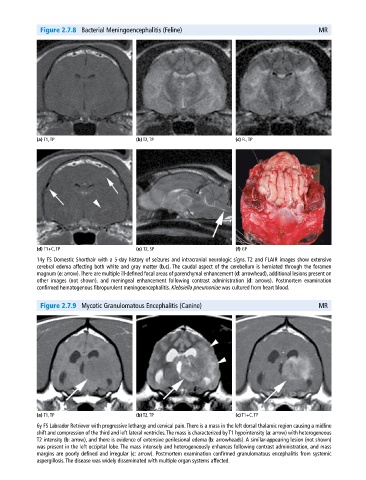
Figure 2.7.8  Bacterial Meningoencephalitis (Feline)                                        MR


             (a) T1, TP                       (b) T2, TP                       (c) FL, TP


             (d) T1+C, TP                     (e) T2, SP                       (f) GP
             14y FS Domestic Shorthair with a 5‐day history of seizures and intracranial neurologic signs. T2 and FLAIR images show extensive
               cerebral edema affecting both white and gray matter (b,c). The caudal aspect of the cerebellum is herniated through the foramen
               magnum (e: arrow). There are multiple ill‐defined focal areas of parenchymal enhancement (d: arrowhead), additional lesions present on
             other images (not shown), and meningeal enhancement following contrast administration (d: arrows). Postmortem examination
               confirmed hematogenous fibropurulent meningoencephalitis. Klebsiella pneumoniae was cultured from heart blood.


              Figure 2.7.9  Mycotic Granulomatous Encephalitis (Canine)                                   MR


             (a) T1, TP                       (b) T2, TP                       (c) T1+C, TP
             6y FS Labrador Retriever with progressive lethargy and cervical pain. There is a mass in the left dorsal thalamic region causing a midline
             shift and compression of the third and left lateral ventricles. The mass is characterized by T1 hypointensity (a: arrow) with heterogeneous
             T2 intensity (b: arrow), and there is evidence of extensive perilesional edema (b: arrowheads). A similar‐appearing lesion (not shown)
             was present in the left occipital lobe. The mass intensely and heterogeneously enhances following contrast administration, and mass
             margins are poorly defined and irregular (c: arrow). Postmortem examination confirmed granulomatous encephalitis from systemic
             aspergillosis. The disease was widely disseminated with multiple organ systems affected.