
Page 223 - Atlas of Small Animal CT and MRI
P. 223
Infectious Inflammatory Disorders 213
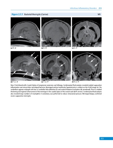
Figure 2.7.7 Bacterial Meningitis (Canine) MR
(a) T1, SP (b) T1, TP (c) FL, TP
(d) T1+C, SP (e) T1+C, TP (f) T1+C, TP
9mo F Dachshund with 2‐week history of progressive weakness and lethargy. Cerebrospinal fluid analysis revealed marked suppurative
inflammation and intracellular rod‐shaped bacteria. Meningeal and periventricular hyperintensity is evident on the FLAIR image (c). The
cerebellum appears enlarged with loss of cerebellar folia definition (c) and ventral foraminal herniation (d: arrowhead). There is marked
thickening and regional enhancement of the meninges in the caudal aspect of the cranial vault (d–f: arrows). Cerebrospinal fluid analysis
also revealed large numbers of neutrophils. A craniotomy was performed to reduce intracranial pressure. Meningeal biopsy confirmed
severe suppurative meningitis.
213