
Page 222 - Atlas of Small Animal CT and MRI
P. 222
212 Atlas of Small Animal CT and MRI
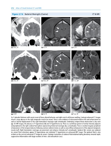
Figure 2.7.6 Bacterial Meningitis (Canine) CT & MR
(a) CT+C, TP (b) T1, TP (c) T2, TP
(d) CT+C, TP (e) T1+C, TP (f) T1+C, DP
(g) CT, TP (h) T2, TP (i) T1+C, TP
3y F Labrador Retriever with recent onset of fever, altered behavior, and right cranial soft‐tissue swelling. Contrast‐enhanced CT images
reveal a large abscess in the right temporalis muscle (a: arrow). There is also evidence of intracranial midline shift and enhancement as
well as internal displacement of the right hemispheric meninges (a,d: arrowheads). Underlying temporal bone destruction is also seen
(g). On MR images, the abscess is T1 hypointense (b) and T2 hyperintense (c). There is a curvilinear pattern of similar intensity adjacent
and peripheral to the right cerebral hemisphere representing thickened meninges (b,c: arrowheads). There is marked irregular peripheral
enhancement of the temporal abscess following contrast administration as well as more diffuse enhancement of the adjacent temporalis
muscle (e,f). Right hemispheric meninges are prominent and enhance intensely (e,f: arrowheads). Epidural (h,i: arrow) and subdural
(e: arrow) fluid collections appear T2 hyperintense and relatively T1 hypointense on enhanced MR images. The epidural fluid is also
evident on the contrast‐enhanced CT image (d: arrow). Cytologic evaluation of subdural fluid obtained during durotomy revealed septic
suppurative inflammation with large numbers of intra‐ and extracellular cocci.