
Page 218 - Atlas of Small Animal CT and MRI
P. 218
208 Atlas of Small Animal CT and MRI
cerebellitis with subsequent cerebellar atrophy has been and T2 signal intensity will vary depending on the age of
linked to Neospora caninum infection (Figure 2.7.14), the hemorrhage and is hypointense on T2* gradient echo
and a report of two canine patients with CNS leishma- sequences because of susceptibility effects. 28–31 MR imag-
niasis describes MR features consistent with multiple ing features consistent with meningitis have also been
nonhemorrhagic infarcts. 24,25 described with this disorder. 30
Helminth‐induced meningoencephalopathy Neurocysticercosis
Neurocysticercosis is a rare form of inflammatory
Angiostrongylus vasorum brain disease caused by aberrant larval migration and
There are multiple reports of Angiostrongylus vasorum, the development of Taenia crassiceps. One MR imaging
French heartworm, producing vasculitis and coagulopathy report of neurocysticercosis in a dog describes cyst‐like
that lead to hemorrhagic inflammatory brain disease in lesions in the subdural region of one occipital lobe and
dogs. Lesions include multifocal brain hemorrhages that the brainstem. Cyst margins enhanced on contrast‐
may include large space‐occupying hematomas. Lesion T1 enhanced T1 images. 32
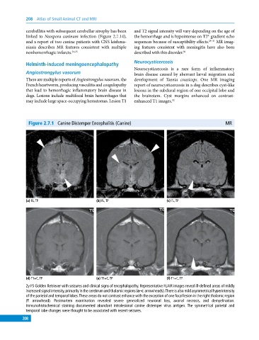
Figure 2.7.1 Canine Distemper Encephalitis (Canine) MR
(a) FL, TP (b) FL, TP (c) FL, TP
(d) T1+C, TP (e) T1+C, TP (f) T1+C, TP
2y FS Golden Retriever with seizures and clinical signs of encephalopathy. Representative FLAIR images reveal ill‐defined areas of mildly
increased signal intensity, primarily in the cerebrum and thalamic regions (a–c: arrowheads). There is also mild asymmetrical hyperintensity
of the parietal and temporal lobes. These areas do not contrast enhance with the exception of one focal lesion in the right thalamic region
(f: arrowhead). Postmortem examination revealed severe generalized neuronal loss, axonal necrosis, and demyelination.
Immunohistochemical staining documented abundant intralesional canine distemper virus antigen. The symmetrical parietal and
temporal lobe changes were thought to be associated with recent seizures.
208