
Page 214 - Atlas of Small Animal CT and MRI
P. 214
204 Atlas of Small Animal CT and MRI
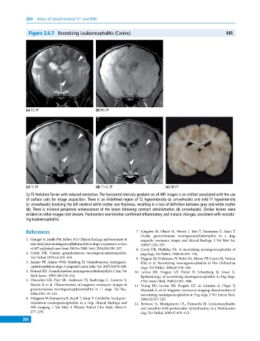
Figure 2.6.7 Necrotizing Leukoencephalitis (Canine) MR
(a) T2, TP (b) PD, TP
(c) T1, TP (d) T1+C, TP (e) GP, TP
2y FS Yorkshire Terrier with reduced mentation. The horizontal intensity gradient on all MR images is an artifact associated with the use
of surface coils for image acquisition. There is an ill‐defined region of T2 hyperintensity (a: arrowheads) and mild T1 hypointensity
(c: arrowheads) involving the left cerebral white matter and thalamus, resulting in a loss of definition between gray and white matter
(b). There is minimal peripheral enhancement of the lesion following contrast administration (d: arrowheads). Similar lesions were
evident on other images (not shown). Postmortem examination confirmed inflammatory and malacic changes, consistent with necrotiz
ing leukoencephalitis.
References 7. Kitagawa M, Okada M, Watari T, Sato T, Kanayama K, Sakai T.
Ocular granulomatous meningoencephalomyelitis in a dog:
1. Granger N, Smith PM, Jeffery ND. Clinical findings and treatment of magnetic resonance images and clinical findings. J Vet Med Sci.
non‐infectious meningoencephalomyelitis in dogs: a systematic review 2009;71:233–237.
of 457 published cases from 1962 to 2008. Vet J. 2010;184:290–297. 8. Cordy DR, Holliday TA. A necrotizing meningoencephalitis of
2. Cordy DR. Canine granulomatous meningoencephalomyelitis. pug dogs. Vet Pathol. 1989;26:191–194.
Vet Pathol. 1979;16:325–333. 9. Higgins RJ, Dickinson PJ, Kube SA, Moore PF, Couto SS, Vernau
3. Adamo PF, Adams WM, Steinberg H. Granulomatous meningoen- KM, et al. Necrotizing meningoencephalitis in five Chihuahua
cephalomyelitis in dogs. Compend Contin Educ Vet. 2007;29:678–690. dogs. Vet Pathol. 2008;45:336–346.
4. Braund KG. Granulomatous meningoencephalomyelitis. J Am Vet 10. Levine JM, Fosgate GT, Porter B, Schatzberg SJ, Greer K.
Med Assoc. 1985;186:138–141. Epidemiology of necrotizing meningoencephalitis in Pug dogs.
5. Cherubini GB, Platt SR, Anderson TJ, Rusbridge C, Lorenzo V, J Vet Intern Med. 2008;22:961–968.
Mantis P, et al. Characteristics of magnetic resonance images of 11. Young BD, Levine JM, Fosgate GT, de Lahunta A, Flegel T,
granulomatous meningoencephalomyelitis in 11 dogs. Vet Rec. Matiasek K, et al. Magnetic resonance imaging characteristics of
2006;159:110–115. necrotizing meningoencephalitis in Pug dogs. J Vet Intern Med.
6. Kitagawa M, Kanayama K, Satoh T, Sakai T. Cerebellar focal gran- 2009;23:527–535.
ulomatous meningoencephalitis in a dog: clinical findings and 12. Berrocal A, Montgomery DL, Pumarola M. Leukoencephalitis
MR imaging. J Vet Med A Physiol Pathol Clin Med. 2004;51: and vasculitis with perivascular demyelination in a Weimaraner
277–279. dog. Vet Pathol. 2000;37:470–472.
204