
Page 212 - Atlas of Small Animal CT and MRI
P. 212
202 Atlas of Small Animal CT and MRI
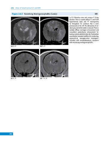
Figure 2.6.5 Necrotizing Meningoencephalitis (Canine) MR
1y FS Chihuahua cross with ataxia of 10‐day
duration. There is marked, diffuse T2 and FLAIR
hyperintensity (a,b) and T1 heterogeneity
(c) throughout the cerebrum that is more
pronounced on the left. The delineation of cer
ebral gray and white matter is reduced. There is
marked, diffuse meningeal enhancement and
nonuniform parenchymal enhancement fol
lowing contrast administration (d). Postmortem
examination revealed multifocal to coalescing,
asymmetrical, nonsuppurative meningoen
cephalitis with encephalomalacia, consistent
with necrotizing meningoencephalitis.
(a) T2, TP (b) FL, TP
(c) T1, TP (d) T1+C, TP
202