
Page 210 - Atlas of Small Animal CT and MRI
P. 210
200 Atlas of Small Animal CT and MRI
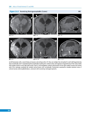
Figure 2.6.3 Necrotizing Meningoencephalitis (Canine) MR
(a) T2, TP (b) T1, TP (c) T1, SP
(d) FL, TP (e) T1+C, TP (f) T1+C, SP
3y M Pomeranian with a recent history of seizures and circling to the left. There are multiple foci of marked T2 and FLAIR hyperintensity
(a,d: arrowheads) and T1 hypointensity (b: arrowheads) involving the cerebral cortex and regional T2 hyperintensity of the white matter
and caudate nucleus on the right (a,d: arrow). There is mild amorphous contrast enhancement of the right caudate nucleus (e,f: arrow)
and of the meninges overlying the cerebral cortical lesions (e,f: arrowheads). Postmortem examination revealed extensive areas of
cortical necrosis and encephalomalacia, consistent with necrotizing meningoencephalitis.
200