
Page 211 - Atlas of Small Animal CT and MRI
P. 211
Noninfectious Inflammatory Disorders 201
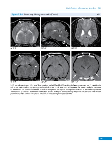
Figure 2.6.4 Necrotizing Meningoencephalitis (Canine) MR
(a) T2, TP (b) T2, SP (c) T2, DP
(d) FL, TP (e) T1, TP (f) T1+C, TP
2y FS Pug with recent onset of lethargy. There is regional marked T2 and FLAIR hyperintensity (a,c,d: arrowheads) and T1 hypointensity
(e,f: arrowheads) involving the frontoparietal cerebral cortex. Uncal (transtentorial) herniation (b: arrow), cerebellar herniation
(b: arrowhead), and interstitial edema (d: arrows) are also present. Widespread meningeal enhancement is seen following contrast
administration (f: arrows). Postmortem examination revealed meningitis and necrotizing encephalitis of gray and white matter
predominately in the cerebral hemispheres, consistent with necrotizing meningoencephalitis.
201