
Page 208 - Atlas of Small Animal CT and MRI
P. 208
198 Atlas of Small Animal CT and MRI
gray and white matter, and typically have indistinct cells, and frank necrosis. Descriptions of the anatomic
15
margins. About half to two thirds of lesions contrast distribution of this disorder are sparse, but lesions can be
enhance on MR, but enhancement is minimal to focal, asymmetrically multifocal, or regionally diffuse
moderate and nonuniform, when present. Meningeal with a predilection for the cerebral hemispheres, although
enhancement is evident in about 50% of patients brainstem lesions have also been reported. 15,16
(Figures 2.6.3, 2.6.4, 2.6.5). 11 Lesions are iso‐ to hypoattenuating on unenhanced
CT images and may appear contiguous with the ventri-
Necrotizing leukoencephalitis cles. Contrast enhancement is absent to moderate and
16
Necrotizing leukoencephalitis is also a nonsuppurative, nonuniform and ill defined, if present. On MR images,
necrotizing, inflammatory brain disorder affecting both brain lesions are T1 hypointense and T2 hyperintense
gray and white matter. 12–15 Grossly, there are subcortical and minimally to moderately contrast enhance. When
regions of liquefaction and cavitation. Microscopically, enhancement is present, it is typically nonuniform and
lesions are characterized by mononuclear infiltrates, gitter sometimes peripheral (Figures 2.6.6, 2.6.7). 15
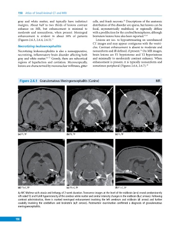
Figure 2.6.1 Granulomatous Meningoencephalitis (Canine) MR
(a) T1, TP (b) T2, TP (c) FL, TP
(d) T1+C, TP (e) T1+C, TP (f) T1+C, SP
4y MC Maltese with ataxia and lethargy of 2‐week duration. Transverse images at the level of the midbrain (a–c) reveal predominantly
left‐sided T2 and FLAIR hyperintensity of the cerebral white matter and similar intensity changes in the midbrain (b,c: arrows). Following
contrast administration, there is marked meningeal enhancement involving the left cerebrum and midbrain (d: arrow) and further
caudally involving the cerebellum and brainstem (e,f: arrows). Postmortem examination confirmed a diagnosis of granulomatous
meningoencephalitis.
198