
Page 213 - Atlas of Small Animal CT and MRI
P. 213
Noninfectious Inflammatory Disorders 203
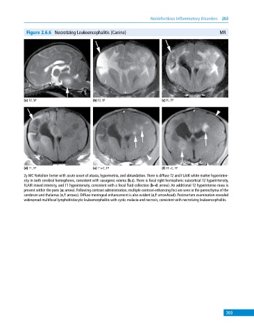
Figure 2.6.6 Necrotizing Leukoencephalitis (Canine) MR
(a) T2, SP (b) T2, TP (c) FL, TP
(d) T1, TP (e) T1+C, TP (f) T1+C, TP
2y MC Yorkshire Terrier with acute onset of ataxia, hypermetria, and obtundation. There is diffuse T2 and FLAIR white matter hyperinten
sity in both cerebral hemispheres, consistent with vasogenic edema (b,c). There is focal right hemispheric subcortical T2 hyperintensity,
FLAIR mixed intensity, and T1 hypointensity, consistent with a focal fluid collection (b–d: arrow). An additional T2 hyperintense mass is
present within the pons (a: arrow). Following contrast administration, multiple contrast‐enhancing foci are seen in the parenchyma of the
cerebrum and thalamus (e,f: arrows). Diffuse meningeal enhancement is also evident (e,f: arrowhead). Postmortem examination revealed
widespread multifocal lymphohistiocytic leukoencephalitis with cystic malacia and necrosis, consistent with necrotizing leukoencephalitis.
203