
Page 209 - Atlas of Small Animal CT and MRI
P. 209
Noninfectious Inflammatory Disorders 199
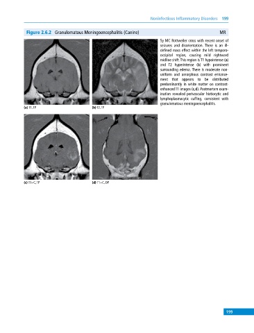
Figure 2.6.2 Granulomatous Meningoencephalitis (Canine) MR
5y MC Rottweiler cross with recent onset of
seizures and disorientation. There is an ill‐
defined mass effect within the left temporo‐
occipital region, causing mild rightward
midline shift. This region is T1 hypointense (a)
and T2 hyperintense (b) with prominent
surrounding edema. There is moderate non
uniform and amorphous contrast enhance
ment that appears to be distributed
predominantly in white matter on contrast‐
enhanced T1 images (c,d). Postmortem exam
ination revealed perivascular histiocytic and
lymphoplasmacytic cuffing, consistent with
granulomatous meningoencephalitis.
(a) T1, TP (b) T2, TP
(c) T1+C, TP (d) T1+C, DP
199