
Page 220 - Atlas of Small Animal CT and MRI
P. 220
210 Atlas of Small Animal CT and MRI
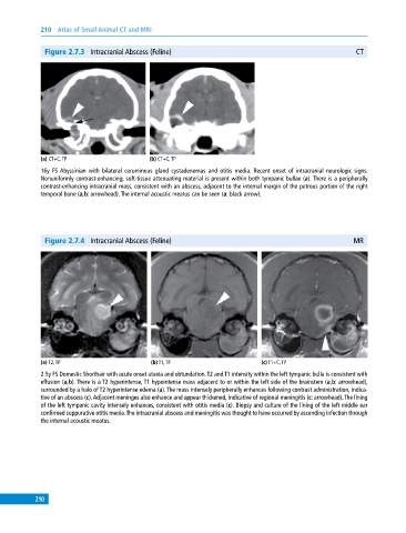
Figure 2.7.3 Intracranial Abscess (Feline) CT
(a) CT+C, TP (b) CT+C, TP
16y FS Abyssinian with bilateral ceruminous gland cystadenomas and otitis media. Recent onset of intracranial neurologic signs.
Nonuniformly contrast‐enhancing, soft‐tissue attenuating material is present within both tympanic bullae (a). There is a peripherally
contrast‐enhancing intracranial mass, consistent with an abscess, adjacent to the internal margin of the petrous portion of the right
temporal bone (a,b: arrowhead). The internal acoustic meatus can be seen (a: black arrow).
Figure 2.7.4 Intracranial Abscess (Feline) MR
(a) T2, TP (b) T1, TP (c) T1+C, TP
2.5y FS Domestic Shorthair with acute onset ataxia and obtundation. T2 and T1 intensity within the left tympanic bulla is consistent with
effusion (a,b). There is a T2 hyperintense, T1 hypointense mass adjacent to or within the left side of the brainstem (a,b: arrowhead),
surrounded by a halo of T2 hyperintense edema (a). The mass intensely peripherally enhances following contrast administration, indica-
tive of an abscess (c). Adjacent meninges also enhance and appear thickened, indicative of regional meningitis (c: arrowhead). The lining
of the left tympanic cavity intensely enhances, consistent with otitis media (c). Biopsy and culture of the lining of the left middle ear
confirmed suppurative otitis media. The intracranial abscess and meningitis was thought to have occurred by ascending infection through
the internal acoustic meatus.
210