
Page 241 - Atlas of Small Animal CT and MRI
P. 241
Neoplasia 231
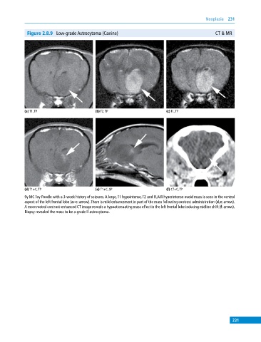
Figure 2.8.9 Low‐grade Astrocytoma (Canine) CT & MR
(a) T1, TP (b) T2, TP (c) FL, TP
(d) T1+C, TP (e) T1+C, SP (f) CT+C, TP
9y MC Toy Poodle with a 3‐week history of seizures. A large, T1 hypointense, T2 and FLAIR hyperintense ovoid mass is seen in the ventral
aspect of the left frontal lobe (a–c: arrow). There is mild enhancement in part of the mass following contrast administration (d,e: arrow).
A more rostral contrast‐enhanced CT image reveals a hypoattenuating mass effect in the left frontal lobe inducing midline shift (f: arrow).
Biopsy revealed the mass to be a grade II astrocytoma.
231