
Page 242 - Atlas of Small Animal CT and MRI
P. 242
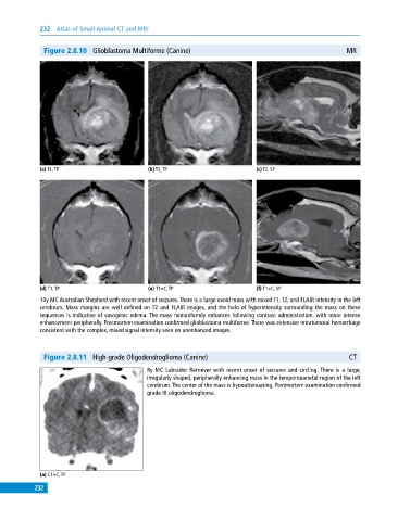
232  Atlas of Small Animal CT and MRI


              Figure 2.8.10  Glioblastoma Multiforme (Canine)                                             MR


             (a) FL, TP                       (b) T2, TP                       (c) T2, SP


             (d) T1, TP                       (e) T1+C, TP                     (f) T1+C, SP
             10y MC Australian Shepherd with recent onset of seizures. There is a large ovoid mass with mixed T1, T2, and FLAIR intensity in the left
             cerebrum. Mass margins are well defined on T2 and FLAIR images, and the halo of hyperintensity surrounding the mass on these
             sequences is indicative of vasogenic edema. The mass nonuniformly enhances following contrast administration, with more intense
             enhancement peripherally. Postmortem examination confirmed glioblastoma multiforme. There was extensive intratumoral hemorrhage
             consistent with the complex, mixed signal intensity seen on unenhanced images.


              Figure 2.8.11  High‐grade Oligodendroglioma (Canine)                                         CT

                                              8y MC Labrador Retriever with recent onset of seizures and circling. There is a large,
                                              irregularly shaped, peripherally enhancing mass in the temporoparietal region of the left
                                              cerebrum. The center of the mass is hypoattenuating. Postmortem examination confirmed
                                              grade III oligodendroglioma.


             (a) CT+C, TP
           232