Page 148 - YORAM RUDY BOOK FINAL
P. 148
P. 148
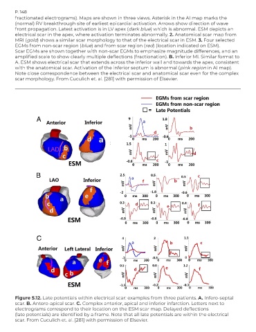
        fractionated electrograms). Maps are shown in three views. Asterisk in the AI map marks the
        (normal) RV breakthrough site of earliest epicardial activation. Arrows show direction of wave
        front propagation. Latest activation is in LV apex (dark blue) which is abnormal. ESM depicts an
        electrical scar in the apex, where activation terminates abnormally. 2. Anatomical scar map from
        MRI (gold) shows a similar scar morphology to that of the electrical scar in ESM. 3. Four selected
        EGMs from non-scar region (blue) and from scar region (red) (location indicated on ESM).
        Scar EGMs are shown together with non-scar EGMs to emphasize magnitude differences, and an
        amplified scale to show clearly multiple deflections (fractionation). B. Inferior MI. Similar format to
        A. ESM shows electrical scar that extends across the inferior wall and towards the apex, consistent
        with the anatomical scar. Activation of the inferior septum is abnormal (pink region in AI map).
        Note close correspondence between the electrical scar and anatomical scar even for the complex
        scar morphology. From Cuculich et. al. [281] with permission of Elsevier.


        Figure 5.12. Late potentials within electrical scar: examples from three patients. A. Infero-septal
        scar. B. Antero-apical scar. C. Complex anterior, apical and inferior infarction. Letters next to
        electrograms correspond to their location on the ESM scar map. Delayed deflections
        (late potentials) are identified by a frame. Note that all late potentials are within the electrical
        scar. From Cuculich et. al. [281] with permission of Elsevier.

