Page 150 - YORAM RUDY BOOK FINAL
P. 150
P. 150
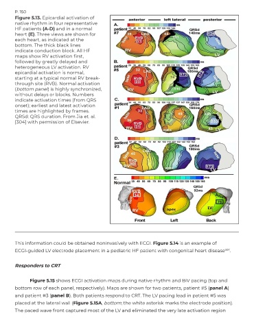
        Figure 5.13. Epicardial activation of
        native rhythm in four representative
        HF patients (A-D) and in a normal
        heart (E). Three views are shown for
        each heart, as indicated at the
        bottom. The thick black lines
        indicate conduction block. All HF
        maps show RV activation first,
        followed by greatly delayed and
        heterogeneous LV activation. RV
        epicardial activation is normal,
        starting at a typical normal RV break-
        through site (RVB). Normal activation
        (bottom panel) is highly synchronized,
        without delays or blocks. Numbers
        indicate activation times (from QRS
        onset); earliest and latest activation
        times are highlighted by frames.
        QRSd: QRS duration. From Jia et. al.
        [304] with permission of Elsevier.


        This information could be obtained noninvasively with ECGI. Figure 5.14 is an example of
        ECGI-guided LV electrode placement in a pediatric HF patient with congenital heart disease           307 .


        Responders to CRT


               Figure 5.15 shows ECGI activation maps during native rhythm and BiV pacing (top and
        bottom row of each panel, respectively). Maps are shown for two patients, patient #5 (panel A)
        and patient #3 (panel B). Both patients respond to CRT. The LV pacing lead in patient #5 was
        placed at the lateral wall (Figure 5.15A, bottom; the white asterisk marks the electrode position).
        The paced wave front captured most of the LV and eliminated the very late activation region

