Page 155 - YORAM RUDY BOOK FINAL
P. 155
P. 155
        deviation of activation times at 500 sites on the LV epicardium. The control value of ED in the

        normal population is 20±4 ms. We defined the LV to be dyssychronous if ED exceeded 28 ms.
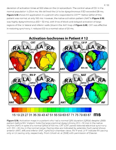
        Figure 5.18 shows ED application in a patient who responded to CRT         308 . Native QRSd of this
        patient was normal, at only 100 ms. However, the native activation pattern (NAT in Figure 5.18)
        was highly dyssynchronous (ED = 32 ms), with lines of block and delayed activation of large

        regions of the LV lateral and inferior walls (blue in the NAT map of Figure 5.18). CRT was effective
        in restoring synchrony; it reduced ED to a normal value of 20 ms.


        Figure 5.18. Activation maps in a patient who had a normal QRS duration (QRSd) despite LBBB
        pattern before BiV implant. Note the large electrical dyssynchrony (ED = 32 ms) in the native
        rhythm (NAT) in spite of a normal QRSd of 100 ms. CRT (CRT panel) restored electrical synchrony
        with ED = 20 ms, in the normal range. Pacing sites are indicated by asterisks. Each panel shows
        anterior (ANT, left) and inferior (INF, right) four-chamber views. RV-P and LV-P indicate RV pacing
        only or LV pacing only, respectively. From Ghosh et. al. [308] with permission of Elsevier.

