Page 159 - YORAM RUDY BOOK FINAL
P. 159
P. 159


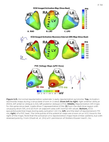
                                        PVC Voltage Maps (LPO View)


        Figure 5.21. Abnormal repolarization substrate in early repolarization syndrome. Top, Activation
        isochrones maps during a sinus beat shown in 3 views (from left to right: right anterior oblique
        (RAO), left anterior oblique (LAO), left posterior oblique (LPO)). Middle, Repolarization ARI maps
        for the same sinus beat. Insets show 2 representative EGMs: (1) from the dark blue region with
        unusually short ARI, and (2) from an adjacent area with normal ARI values. Bottom, ECGI
        epicardial potential maps during early activation (40 ms, left) and the start of repolarization) 170
        ms, right) of a PVC beat. The time points of the potential maps are shown on an ECG trace to the
        right of the maps. Note that the activation and repolarization maps have similar patterns, but with
        reversed polarity. From Ghosh et. al. [314] with permission of Wolters Kluwer Health, Inc.

