Page 164 - YORAM RUDY BOOK FINAL
P. 164
P. 164


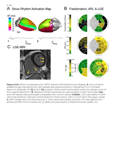
        Figure 5.24. Electric substrate of an ARVC patient with biventricular disease. A. Sinus rhythm
        breakthrough was abnormal, with earliest epicardial activation originating from the basal
        lateral LV (asterisk). RV (1) and LV (2) unipolar EGMs were fractionated (note the voltage scale of
        low-magnitude EGM 1). B. Regions of high fractionation were present in both ventricles (top),
        and ARI values were prolonged compared with control values (middle). LGE was visible in both
        ventricles (bottom). Normal control levels for fractionation, ARI and LGE fall in the green range.
        C. MRI image with LGE showing scar in both ventricles (yellow arrows). LGE: late gadolinium
        enhanced MRI. From Andrews et. al. [323] with permission of Wolters Kluwer Health, Inc.

