Page 169 - YORAM RUDY BOOK FINAL
P. 169
P. 169


          A


         B                                                                                                      C


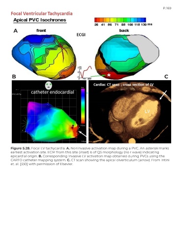
        Figure 5.28. Focal LV tachycardia. A. Noninvasive activation map during a PVC. An asterisk marks
        earliest activation site. EGM from this site (inset) is of QS morphology (no r wave) indicating
        epicardial origin. B. Corresponding invasive LV activation map obtained during PVCs using the
        CARTO catheter mapping system. C. CT scan showing the apical diverticulum (arrow). From Intini
        et. al. [330] with permission of Elsevier.

