Page 171 - YORAM RUDY BOOK FINAL
P. 171
P. 171
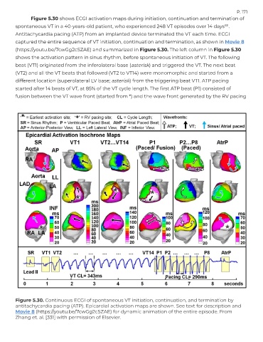
               Figure 5.30 shows ECGI activation maps during initiation, continuation and termination of

        spontaneous VT in a 40-years-old patient, who experienced 248 VT episodes over 14 days .
                                                                                                         331
        Antitachycardia pacing (ATP) from an implanted device terminated the VT each time. ECGI
        captured the entire sequence of VT initiation, continuation and termination, as shown in Movie 8
        (https://youtu.be/7cwGg2cSZAE) and summarized in Figure 5.30. The left column in Figure 5.30

        shows the activation pattern in sinus rhythm, before spontaneous initiation of VT. The following
        beat (VT1) originated from the inferolateral base (asterisk) and triggered the VT. The next beat
        (VT2) and all the VT beats that followed (VT2 to VT14) were monomorphic and started from a
        different location (superolateral LV base; asterisk) from the triggering beat VT1. ATP pacing

        started after 14 beats of VT, at 85% of the VT cycle length. The first ATP beat (P1) consisted of
        fusion between the VT wave front (started from *) and the wave front generated by the RV pacing


        Figure 5.30. Continuous ECGI of spontaneous VT initiation, continuation, and termination by
        antitachycardia pacing (ATP). Epicardial activation maps are shown. See text for description and
        Movie 8 (https://youtu.be/7cwGg2cSZAE) for dynamic animation of the entire episode. From
        Zhang et. al. [331] with permission of Elsevier.

