Page 174 - YORAM RUDY BOOK FINAL
P. 174
P. 174
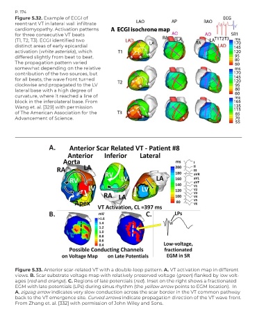
        Figure 5.32. Example of ECGI of
        reentrant VT in lateral wall infiltrate
        cardiomyopathy. Activation patterns
        for three consecutive VT beats
        (T1, T2, T3). ECGI identified two
        distinct areas of early epicardial
        activation (white asterisks), which
        differed slightly from beat to beat.
        The propagation pattern varied
        somewhat depending on the relative
        contribution of the two sources, but
        for all beats, the wave front turned
        clockwise and propagated to the LV
        lateral base with a high degree of
        curvature, where it reached a line of
        block in the inferolateral base. From
        Wang et. al. [329] with permission
        of The American Association for the
        Advancement of Science.


                        A.


                        B.                                             C.


        Figure 5.33. Anterior scar-related VT with a double-loop pattern. A. VT activation map in different
        views. B. Scar substrate voltage map with relatively preserved voltage (green) flanked by low volt-
        ages (red and orange). C. Regions of late potentials (red). Inset on the right shows a fractionated
        EGM with late potentials (LPs) during sinus rhythm (the yellow arrow points to EGM location).  In
        A, zigzag arrow indicates very slow conduction across the scar border in the VT common pathway
        back to the VT emergence site. Curved arrows indicate propagation direction of the VT wave front.
        From Zhang et. al. [332] with permission of John Wiley and Sons.

