Page 170 - YORAM RUDY BOOK FINAL
P. 170
P. 170


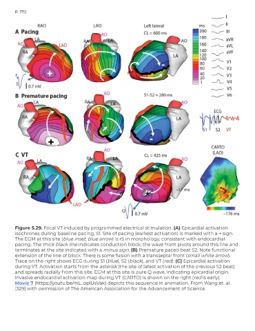
        Figure 5.29. Focal VT induced by programmed electrical stimulation. (A) Epicardial activation
        isochrones during baseline pacing, S1. Site of pacing (earliest activation) is marked with a + sign.
        The EGM at this site (blue inset; blue arrow) is rS in morphology, consistent with endocardial
        pacing. The thick black line indicates conduction block; the wave front pivots around this line and
        terminates at the site indicated with a minus sign. (B) Premature paced beat S2. Note functional
        extension of the line of block. There is some fusion with a transseptal front (small white arrow).
        Trace on the right shows ECG during S1 (blue), S2 (black), and VT (red). (C) Epicardial activation
        during VT. Activation starts from the asterisk (the site of latest activation of the previous S2 beat)
        and spreads radially from this site. EGM at this site is pure Q wave, indicating epicardial origin.
        Invasive endocardial activation map during VT (CARTO) is shown on the right (red is early).
        Movie 7 (https://youtu.be/mL_oplUsVak) depicts this sequence in animation. From Wang et. al.
        [329] with permission of The American Association for the Advancement of Science.

