Page 168 - YORAM RUDY BOOK FINAL
P. 168
P. 168


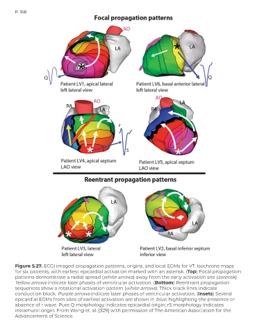
        Figure 5.27. ECGI imaged propagation patterns, origins, and local EGMs for VT. Isochrone maps
        for six patients, with earliest epicardial activation marked with an asterisk. (Top) Focal propagation
        patterns demonstrate a radial spread (white arrows) away from the early activation site (asterisk).
        Yellow arrows indicate later phases of ventricular activation. (Bottom) Reentrant propagation
        sequences show a rotational activation pattern (white arrows). Thick black lines indicate
        conduction block. Purple arrows indicate later phases of ventricular activation. (Insets) Several
        epicardial EGMs from sites of earliest activation are shown in blue, highlighting the presence or
        absence of r wave. Pure Q morphology indicates epicardial origin; rS morphology indicates
        intramural origin. From Wang et. al. [329] with permission of The American Association for the
        Advancement of Science.

