Page 167 - YORAM RUDY BOOK FINAL
P. 167
P. 167


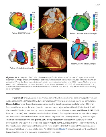
        Figure 5.26. Examples of ECGI isochrones maps for localization of VT site of origin. Epicardial
        isochrones maps are shown for four patients, with earliest epicardial activation marked with an
        asterisk. EP study-determined sites of origin are indicated below the ECGI maps. Yellow arrows
        point to VT origin on a representative CT scan. From Wang et. al. [329] with permission of The
        American Association for the Advancement of Science. AO, aorta; LAD, left anterior descending
        coronary artery


               Figure 5.29 shows an example from a patient with nonischemic cardiomyopathy              329 . ECGI
        was applied in the EP laboratory during induction of VT by programmed electrical stimulation.
        Figure 5.29A shows the activation sequence during baseline pacing (cycle length = 600 ms)
        from a catheter in the RV apex (location marked by a + sign). A line of block (thick black line) in

        the lateral LV is circumvented by the excitation wave front. Premature pacing (Figure 5.29B; S1-S2
        interval = 280 ms) functionally extends the line of block , forcing the wave front to make a longer
        arc around this line and activate a more inferior region of the LV last (marked by a minus sign).

        The first VT beat is shown in Figure 5.29C; it originates from the location (asterisk) of latest
        activation by the S2 premature paced beat in Figure 5.29B, suggesting that triggered activity is
        the mechanism of initiation of this focal VT. Note that the EGM at the site of VT origin is a pure
        Q-wave, indicating an epicardial origin. An ECGI movie (Movie 7, https://youtu.be/mL_oplUsVak)
        is provided to show the dynamic progression in this case.

