
Page 300 - Clinical Manual of Small Animal Endosurgery
P. 300
288 Clinical Manual of Small Animal Endosurgery
Fig. 10.9 Due to their thin bladder wall, cystoscopy allows a degree of
transmural visualisation of some coelomic organs in chelonians. In this case
the right ovary, which is inactive, and the edge of the liver are evident.
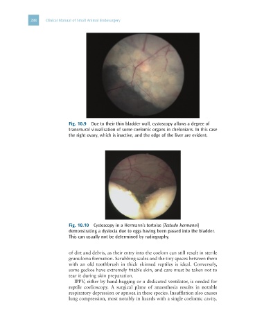
Fig. 10.10 Cystoscopy in a Hermann’s tortoise (Testudo hermanni)
demonstrating a dystocia due to eggs having been passed into the bladder.
This can usually not be determined by radiography.
of dirt and debris, as their entry into the coelom can still result in sterile
granuloma formation. Scrubbing scales and the tiny spaces between them
with an old toothbrush in thick skinned reptiles is ideal. Conversely,
some geckos have extremely friable skin, and care must be taken not to
tear it during skin preparation.
IPPV, either by hand-bagging or a dedicated ventilator, is needed for
reptile coelioscopy. A surgical plane of anaesthesia results in notable
respiratory depression or apnoea in these species. Insufflation also causes
lung compression, most notably in lizards with a single coelomic cavity.