
Page 303 - Clinical Manual of Small Animal Endosurgery
P. 303
Small Exotic Animal Endosurgery 291
biopsies may suffer from crush artefact. The kidneys are extracoelomic,
and in many species may either be difficult to visualise behind the pig-
mented coelomic membrane, or positioned in the pelvis where they
cannot easily be accessed. In those species, such as terrapins, where they
are accessible, it is recommended to incise the overlying coelomic mem-
brane for biopsy access; otherwise samples will suffer from notable crush
artefact.
The saurian lung is a unicameral balloon-like structure, with spongy
faveolar gaseous-exchange tissue, rather than alveoli. The faveolar struc-
ture gradually reduces caudally through the lung, leaving the caudal
section as a relatively avascular thin-walled air sac.
Jekl and Knotek (2006) and Stahl et al. (2008) described the endo-
scopic examination of the inner lung surface in snakes, and Divers (1999)
described a similar technique in green iguanas. This not only allows
visual examination but also sampling for microbiological culture, biop-
sies for histology and removal of pentastomid parasites in the lungs
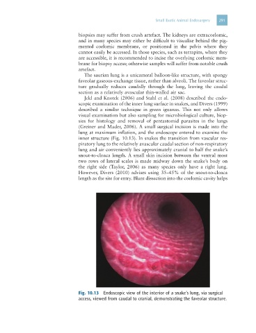
(Greiner and Mader, 2006). A small surgical incision is made into the
lung at maximum inflation, and the endoscope entered to examine the
inner structure (Fig. 10.13). In snakes the transition from vascular res-
piratory lung to the relatively avascular caudal section of non-respiratory
lung and air conveniently lies approximately cranial to half the snake’s
snout-to-cloaca length. A small skin incision between the ventral most
two rows of lateral scales is made midway down the snake’s body on
the right side (Taylor, 2006) as many species only have a right lung.
However, Divers (2010) advises using 35–45% of the snout-to-cloaca
length as the site for entry. Blunt dissection into the coelomic cavity helps
Fig. 10.13 Endoscopic view of the interior of a snake’s lung, via surgical
access, viewed from caudal to cranial, demonstrating the faveolar structure.