
Page 307 - Clinical Manual of Small Animal Endosurgery
P. 307
Small Exotic Animal Endosurgery 295
endoscopy-aided intubation is performed, positioning is vital to success.
The rabbit’s neck needs to be held vertically and extended, slightly lifting
the front of the rabbit’s body from the table, to allow dislocation of the
soft palate from above the epiglottis. If this is not performed, intubation,
even if aided with an endoscope, is almost impossible. The rabbit also
needs to have reached a reasonable plane of anaesthesia after induction,
otherwise the strong swallowing reflex makes intubation difficult. Suf-
ficient time is needed after intramuscular induction.
Vaginoscopy
Vaginoscopy as part of the diagnostic work-up for haematurea in rabbits
is unfortunately not usually helpful in determining whether the underly-
ing cause is a uterine adenocarcinoma. The double cervix is usually
unremarkable in appearance and closed. Vaginoscopy is however useful
in determining if a female rabbit has been previously neutered. The
technique is performed under anaesthesia. The endoscope and sheath are
inserted in the vulva and the vagina inflated with saline while the vulva
is pinched closed by the operator’s fingers. Most clinicians will perform
rabbit ovariohysterectomies including the cervix to prevent the risk of
later development of an adenocarcinoma in the remnant uterine tissue.
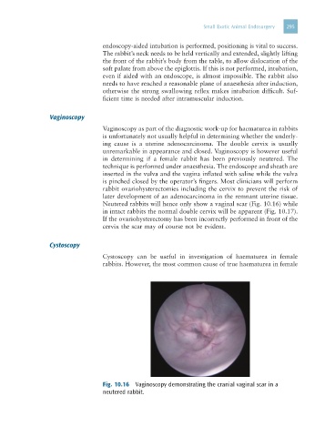
Neutered rabbits will hence only show a vaginal scar (Fig. 10.16) while
in intact rabbits the normal double cervix will be apparent (Fig. 10.17).
If the ovariohysterectomy has been incorrectly performed in front of the
cervix the scar may of course not be evident.
Cystoscopy
Cystoscopy can be useful in investigation of haematurea in female
rabbits. However, the most common cause of true haematurea in female
Fig. 10.16 Vaginoscopy demonstrating the cranial vaginal scar in a
neutered rabbit.