
Page 302 - Clinical Manual of Small Animal Endosurgery
P. 302
290 Clinical Manual of Small Animal Endosurgery
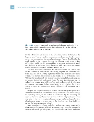
Fig. 10.12 A ventral approach to coelioscopy in lizards, such as for this
liver biopsy, yields reduced access and visualisation due to the midline
abdominal vein, caudal to the liver.
to the pelvis until just cranial to the umbilicus, where it then joins the
hepatic vein. This vein and its suspensory membrane can hinder visuali-
sation and exploration via ventral coelioscopy. Access should either be
made caudal to the junction between the bilateral pelvic veins, or just
off midline, and is usually accomplished by an open technique. A small
skin incision is made and blunt dissection with haemostats performed
until the coelom is entered, and the cannula placed.
Tortoises and other chelonians are ideal candidates for coelioscopy,
as the alternative, transplastral coeliotomy, requires an osteotomy and
bone flap, and has a notably higher morbidity and mortality associated
with it. The normal access site is in the middle of the prefemoral fossa.
While either can be used, it is more ergonomic for right-handed surgeons
to operate in the left prefemoral fossa. As most tortoises can have a
voluminous bladder that will interfere with coelioscopy, it is advisable
to encourage urination before anaesthesia, by stimulating the cloaca.
Access is open, with dissection using a blunt-tipped haemostat as in
lizards.
Despite the simple anatomy of snakes, coelioscopy yields poor visu-
alisation in most cases. Insufflation usually fails to result in any meaning-
ful operating space, as the coelom is constrained by the encircling rib
cage, and snakes also have more diffuse coelomic adipose tissue. There
may also be fibrous connections between organ surfaces. Recently, visu-
alisation and access to organs such as the liver has been described from
across the lung surface (see below).
After visualisation of any pathology and target organs, biopsy tech-
nique is generally routine. Multiple biopsies are recommended, as samples
are typically small, pathology may not be diffusely distributed and some