
Page 301 - Clinical Manual of Small Animal Endosurgery
P. 301
Small Exotic Animal Endosurgery 289
(a) (b)
(c) (d)
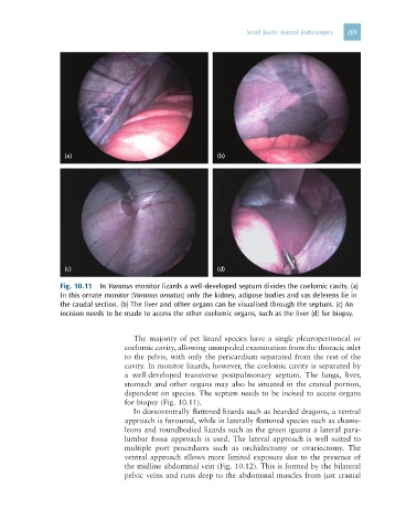
Fig. 10.11 In Varanus monitor lizards a well-developed septum divides the coelomic cavity. (a)
In this ornate monitor (Varanus ornatus) only the kidney, adipose bodies and vas deferens lie in
the caudal section. (b) The liver and other organs can be visualised through the septum. (c) An
incision needs to be made to access the other coelomic organs, such as the liver (d) for biopsy.
The majority of pet lizard species have a single pleuroperitoneal or
coelomic cavity, allowing unimpeded examination from the thoracic inlet
to the pelvis, with only the pericardium separated from the rest of the
cavity. In monitor lizards, however, the coelomic cavity is separated by
a well-developed transverse postpulmonary septum. The lungs, liver,
stomach and other organs may also be situated in the cranial portion,
dependent on species. The septum needs to be incised to access organs
for biopsy (Fig. 10.11).
In dorsoventrally flattened lizards such as bearded dragons, a ventral
approach is favoured, while in laterally flattened species such as chame-
leons and roundbodied lizards such as the green iguana a lateral para-
lumbar fossa approach is used. The lateral approach is well suited to
multiple port procedures such as orchidectomy or ovariectomy. The
ventral approach allows more limited exposure due to the presence of
the midline abdominal vein (Fig. 10.12). This is formed by the bilateral
pelvic veins and runs deep to the abdominal muscles from just cranial