
Page 306 - Clinical Manual of Small Animal Endosurgery
P. 306
294 Clinical Manual of Small Animal Endosurgery
recommended even under anaesthesia. A dental probe is needed to prop-
erly assess any loose teeth or small inapparent diastemas between teeth
that could lead to dental root abscesses. As visualisation is easier with
the oral endoscope there is a reduced risk of post-dental examination
pain and anorexia due to spreading the oral gag too widely, with result-
ant masseter muscle tears or temporomandibular joint injuries. The
endoscope can be periodically inserted during the hand-filing or motor-
ised burring of overgrown molar spurs.
Endoscopy-assisted intubation
The intubation of rabbits can be difficult (as in guinea pigs and chinchil-
las) due to their long narrow mouths and long soft palate that normally
overlies the epiglottis (as obligate nasal breathers). Recommendations
for assisting with intubation include using an otoscope, or intubating
blindly by listening to the rabbit’s respiratory sounds emanating from
the tube. Practice has much to do with ease and success of blind intuba-
tion. Texts advise the use of an endoscope inserted down the endotra-
cheal tube to aid intubation (Harcourt-Brown, 2002), and this can
certainly be useful in difficult cases. There are disadvantages, however.
The endotracheal tube may need to be cut shorter, to allow the endoscope
to reach the end for visualisation. In small rabbits and rodents a 1.9 mm
endoscope is needed to fit within the narrow endotracheal lumen, and
these scopes are very delicate and easily damaged. They can also result
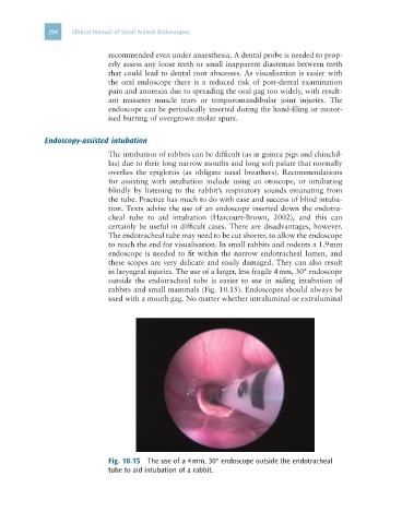
in laryngeal injuries. The use of a larger, less fragile 4 mm, 30° endoscope
outside the endotracheal tube is easier to use in aiding intubation of
rabbits and small mammals (Fig. 10.15). Endoscopes should always be
used with a mouth gag. No matter whether intraluminal or extraluminal
Fig. 10.15 The use of a 4 mm, 30° endoscope outside the endotracheal
tube to aid intubation of a rabbit.