
Page 308 - Clinical Manual of Small Animal Endosurgery
P. 308
296 Clinical Manual of Small Animal Endosurgery
Fig. 10.17 Vaginoscopy demonstrating the normal double cervix in an
intact rabbit.
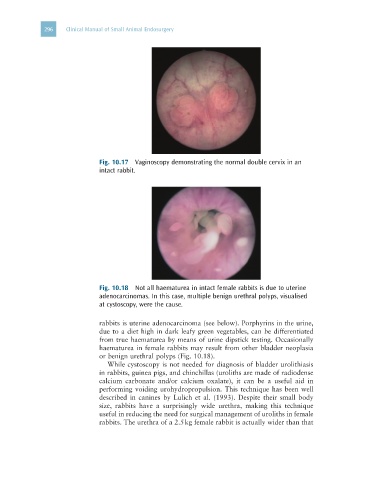
Fig. 10.18 Not all haematurea in intact female rabbits is due to uterine
adenocarcinomas. In this case, multiple benign urethral polyps, visualised
at cystoscopy, were the cause.
rabbits is uterine adenocarcinoma (see below). Porphyrins in the urine,
due to a diet high in dark leafy green vegetables, can be differentiated
from true haematurea by means of urine dipstick testing. Occasionally
haematurea in female rabbits may result from other bladder neoplasia
or benign urethral polyps (Fig. 10.18).
While cystoscopy is not needed for diagnosis of bladder urolithiasis
in rabbits, guinea pigs, and chinchillas (uroliths are made of radiodense
calcium carbonate and/or calcium oxalate), it can be a useful aid in
performing voiding urohydropropulsion. This technique has been well
described in canines by Lulich et al. (1993). Despite their small body
size, rabbits have a surprisingly wide urethra, making this technique
useful in reducing the need for surgical management of uroliths in female
rabbits. The urethra of a 2.5 kg female rabbit is actually wider than that