
Page 47 - Live-cellanalysis handbook
P. 47
Kinetic Cell Migration and Invasion Assays
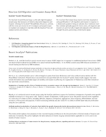
How Live-Cell Migration and Invasion Assays Work
IncuCyte® Scratch Wound Assay IncuCyte® Chemotaxis Assay
The IncuCyte Scratch Wound Assay is a 96-well, high throughput The IncuCyte Chemotaxis Assay enables real-time visualization
wound assay that measures cell density-dependent migration and and quantification of cell migration and invasion in response to
invasion. Utilizing the IncuCyte® WoundMaker, 96 precise, uniform, a chemotactic gradient. Using an optically clear membrane insert
cell-free zones are created in cell monolayers and wound closure that contains optimally spaced 8-micron pores, cell migration and
is visualized and analyzed in real-time with the IncuCyte® live-cell invasion are automatically imaged and analyzed as the cells move
analysis system and software. through the pores, generating a chemotactic signal.
References
1. Cell Migration: integrating signals from front to back. Ridley, A.J., Schwartz, M.A., Burridge, K., Firtel, R.A., Ginsberg, M.H., Borisy, G., Parsons. J.T. and
Horwitz, A.R. Science 302: 1704.
2. Cell Migration and Invasion Assays as Tools for Drug Discovery. Hulkower, K. I and Herber, R. L. Pharmaceutics 2011 3:107
Recent IncuCyte® Publications
Scratch wound assay
Drebert, et. al., used the IncuCyte scratch wound assay to assess HUVEC migration in response to conditioned medium from solvent- (CMS)
and dexamethasone (Dex)-treated (CMD) colon cancer-derived myofibroblasts. In the HUVEC scratch assay CMS-induced acceleration of
wound healing was blunted by CMD treatment.
Colon cancer-derived myofibroblasts increase endothelial cell migration by glucocorticoid-sensitive secretion of a pro-migratory factor. Drebert, Z., MacAskill,
M., Doughty-Shenton, D., De Bosscher, K., Bracke, M., Hadoke, P. W., Beck, I. M. Vascul. Pharmacol., Vascul Pharmacol. 2016 Oct 4. pii: S1537-1891(16)30137-9.
doi: 10.1016/j.vph.2016.10.004. [Epub ahead of print], 2017.
Härmä, et. al., cultured prostate cancer cells on ImageLock plates from Essen BioScience until fully confluent and scratched with the
WoundMaker instrument. Wound closure was monitored and quantified with the IncuCyte® system. Study results suggest that some
betulin-derivatives may specifically target cell motility and invasion by affecting the organization of filamentous actin fiber network at
low nanomolar concentrations.
Optimization of invasion-specific effects of betulin derivatives on prostate cancer cells through lead development. Härmä, V., Haavikko, R., Virtanen,
J., Ahonen, I., Schukov, H. P., Alakurtti, S., Purev, E., Rischer, H., Yli-Kauhaluoma, J., Moreira, V. M., Nees, M., Oksman-Caldentey, K. M. PLoS ONE 05/2015
10(5):e0126111.
Moody, et. al. conducted scratch wound assays using the IncuCyte platform to assess the functional consequence of growth arrest specific
6 (Gas6) neutralization in lung cancer cells. Gas6 is a vitamin-K dependent, 75 kDa growth factor-like protein involved in the regulation
of a wide array of cellular activities, including adhesion, migration, mitogenesis, aggregation, stress-response, differentiation, immune
activation, efferocytosis and apoptosis. A Gas6 neutralizing antibody was also tested for its ability to inhibit Gas6-induced proliferation
using the same platform.
Antibody-mediated neutralization of autocrine Gas6 inhibits the growth of pancreatic ductal adenocarcinoma tumors in vivo. Moody, G., Belmontes,
B., Masterman, S., Wang, W., King, C., Murawsky, C., Tsurda, T., Liu, S., Radinsky, R., Beltran, P. Int J of Cancer. 2016 139:1340.
This review article explores how advances in time-resolved imaging contributes to the characterization of distinct modes of invasion and
molecular mechanisms. The author highlights the latest advances in kinetic imaging instrumentation applicable to in vitro and in vivo
models of tumor invasion.
Profiling distinct mechanisms of tumor invasion for drug discovery: imaging adhesion, signalling and matrix turnover. Carragher, N. Clinical & Exp
Metastasis. 2009 April 26(4).
45