
Page 19 - Rapid Review of ECG Interpretation in Small Animal Practice, 2nd Edition
P. 19
Principles of Electrocardiography
LEAD PLACEMENT AND ACQUISITION • Once the leads are hooked up properly, the ECG
OF THE ECG
leads should not be touching or crossing over
VetBooks.ir The standard ECG recording systems record an • If using ECG clips, it is best to wet the skin and
each other.
analog signal and use a heated stylus which records
on light-sensitive graph paper. Older-style ECG ECG clip with isopropyl alcohol or a coupling
machines recorded one lead at a time while more gel that contains a high ionic concentration to
recent digital systems, which record the ECG on a help aid in the transfer of current at the tissue–
computer, allow any combination of simultaneous electrode interface. If using an “ECG patch”
6- or 12-lead recordings. The ECG machine typi- these often contain gel within the patch and
cally applies a filter to reduce baseline artifact. A alcohol is not required.
50 Hz filter may be adequate in dogs, but in cats • Table 1.1 describes the positive and negative
it is best to use a 150 Hz filter in order to include terminals that form the bipolar and augmented
high-frequency components of the R and S wave. lead system.
Proper grounding is important to avoid electrical
interference from 60 Hz alternating current (AC) Acquisition of the 12-lead ECG:
originating from the electrical supply or other elec-
trical equipment in the room. The ECG reference • Position patient and connect standard limb
ground is provided through a separate electrode, leads as described for 6-lead ECG then attach
either incorporated into one of the three limb leads, the chest leads as follows (Fig. 1.6):
or as a separate wire. • Identify the left 6th intercostal space (IC6 )
L
• Connect V2 just to the left of the sternum at
Acquisition of the 6-lead ECG: IC6 L
• Connect V4 at the costochondral junction of
• Place patient in right lateral recumbency. The IC6 L
animal should have its head and neck resting on • Connect V3 midway between V2 and V4 at
a table or on the floor. IC6 L
• The forelegs and hindlimbs should be parallel • Connect V5 at IC6 at a distance above
L
and at right angles (perpendicular) to the body. V4 equal to the distance between V2–V3 or
• The patient must remain still with minimal V3–V4
panting and moving. Cats should not be • Connect V6 at IC6 at a distance above V5
L
purring. equal to the distance between V2–V3, V3–V4
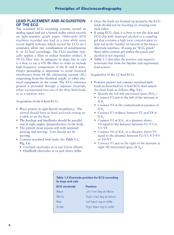
• Connect standard limb leads. See Table 1.2, or V4–V5
Fig. 1.6. • Connect V1 just to the right of the sternum at
• Forelimb electrodes at or just below elbows right 5th intercostal space (IC5 )
R
• Hindlimb electrodes at or just above stifles
Table 1.2 Electrode position for ECG recording
in dogs and cats
ECG electrode Position
Black Left front leg at elbow
White Right front leg at elbow
Red Left back leg at stifle
Green Right back leg at stifle
6