
Page 104 - Fluid, Electrolyte, and Acid-Base Disorders in Small Animal Practice
P. 104
94 ELECTROLYTE DISORDERS
potential. Ionized hypocalcemia increases membrane 80
Stool Potassium Excretion
excitability by allowing self-perpetuating sodium perme-
ability to be reached with a lesser degree of depolariza-
tion, whereas ionized hypercalcemia requires greater 60
than normal depolarization for this threshold to be
reached (see Fig. 5-2). Thus, hypercalcemia counteracts 40
hyperkalemia by normalizing the difference between Percent of daily intake
the resting and threshold potentials, whereas hypocalce-
mia exacerbates the effect of hyperkalemia on membrane 20
excitability. This principle is the basis for treating
hyperkalemia with calcium salts (see the Treatment of
Hyperkalemiasection).Membraneexcitabilityisincreased 0
0 5 10 15 20 25 40 60 80 100
by alkalemia and decreased by acidemia. As aresult of these
Creatinine clearance (mL/min)
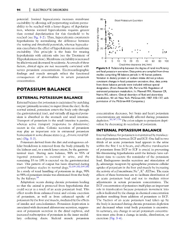
factors, clinical signs are not necessarily correlated with Figure 5-3 Relationship between the degree of renal insufficiency
serum potassium concentrations. Electrocardiographic and fecal potassium excretion. Data points are compiled from three
findings and muscle strength reflect the functional studies comprising 98 balance periods in 40 human patients.
consequences of abnormalities in serum potassium Variation in dietary protein or sodium intake did not produce
concentration. consistent changes in fecal potassium excretion; thus, data points
from these balance periods were included without special
POTASSIUM BALANCE designation. (From Alexander EA, Perrone RD. Regulation of
extrarenal potassium metabolism. In: Maxwell MH, Kleeman CR,
Narins RG, editors. Clinical disorders of fluid and electrolyte
EXTERNAL POTASSIUM BALANCE metabolism, 4th ed. New York: McGraw-Hill, 1987: 105–117, with
permission of the McGraw-Hill Companies.)
External balance for potassium is maintained by matching
output (primarily in urine) to input (from the diet). In the
normal animal, potassium enters the body only through
the gastrointestinal tract, and virtually all ingested potas- concentration decreases, but brain and heart potassium
sium is absorbed in the stomach and small intestine. concentrations are minimally affected during potassium
Transport of potassium in the small intestine is passive, depletion. 20,107,178 The colon adapts to potassium depri-
whereas active transport (responsive to aldosterone) vation by decreasing its secretion of potassium.
occurs in the colon. Colonic secretion of potassium
may play an important role in extrarenal potassium INTERNAL POTASSIUM BALANCE
homeostasis in some disease states (e.g., chronic renal fail- Internal balance for potassium is maintained by transloca-
ure) (Fig. 5-3). tion of potassium between ECF and ICF. One half to two
Potassium derived from the diet and endogenous cel- thirds of an acute potassium load appears in the urine
lular breakdown is removed from the body primarily by within the first 4 to 6 hours, and effective translocation
the kidneys and, to a much lesser extent, by the gastroin- of potassium from ECF to ICF is crucial in preventing
testinal tract. During zero balance, 90% to 95% of life-threatening hyperkalemia until the kidneys have suf-
ingested potassium is excreted in urine, and the ficient time to excrete the remainder of the potassium
remaining 5% to 10% is excreted via the gastrointestinal load. Endogenous insulin secretion and stimulation of
tract. This pattern of output has been observed during b 2 -adrenergic receptors by epinephrine promote cellular
control balance studies in normal dogs. 12,139,152,169,182 uptake of potassium in the liver and muscle by increasing
þ
þ
In a study of renal handling of potassium in dogs, 90% the activity of cell membrane Na ,K -ATPase. The main
to 98% of potassium intake was eliminated from the body effects of these hormones are to facilitate distribution of
by the kidneys. 24 an acute potassium load and not to mediate minor
Adaptation occurs during chronic potassium loading adjustments in serum potassium concentration. The
so that the animal is protected from hyperkalemia that ECF concentration of potassium itself plays an important
could occur as a result of an acute potassium load. This role in translocation because potassium movement into
effect results from enhanced renal and colonic excretion cells is facilitated by the change in chemical concentration
of potassium, as well as from enhanced uptake of gradient resulting from addition of potassium to ECF.
potassium by the liver and muscle, mediated by the effects The fraction of an acute potassium load taken up by
of insulin and catecholamines. Potassium deprivation is the body is increased during chronic potassium depletion
associated with decreased aldosterone secretion, suppres- and decreased when total body potassium is excessive.
sion of potassium secretion in the distal nephron, and In summary, any change in serum potassium concentra-
increased reabsorption of potassium in the inner medul- tion must arise from a change in intake, distribution, or
lary collecting ducts. Skeletal muscle potassium excretion (Fig. 5-4).