
Page 108 - Fluid, Electrolyte, and Acid-Base Disorders in Small Animal Practice
P. 108
98 ELECTROLYTE DISORDERS
extracellular pH play permissive roles in promoting aldo- Interstitial
sterone secretion. Aldosterone release is inhibited by Tubular fluid Cell fluid
dopamine and atrial natriuretic factor, both of which are
released in response to volume expansion.
Slow
Aldosterone increases reabsorption of Na and secre-
þ
tion of K and H ions in the distal nephron. Its primary
þ
þ
Na +
þ
effect is to increase the number of open Na channels in +
3Na
the luminal membranes of the principal cells. Sodium ATP +
2K
reabsorption via these luminal Na channels is electro- K +
þ
genic (i.e., it generates electronegativity in the tubular
lumen). This electronegativity can be dissipated either Luminal flow
by K þ or H þ ion secretion or by Cl reabsorption in
the distal nephron. Aldosterone increases the activity Cl –
þ
þ
and number of Na ,K -ATPase pumps in the basolateral
membranes of the principal cells, and this effect may
þ
occur as a result of increased entry of Na ions across
þ
þ
the luminal membranes. Increased Na ,K -ATPase – +
þ
activity in turn increases the intracellular K concentra-
tion and facilitates K þ secretion across the luminal
Negative Positive
membranes. Aldosterone also increases the number of
open K þ channels in the luminal membrane, thus Fast
facilitating K exit into tubular fluid.
þ
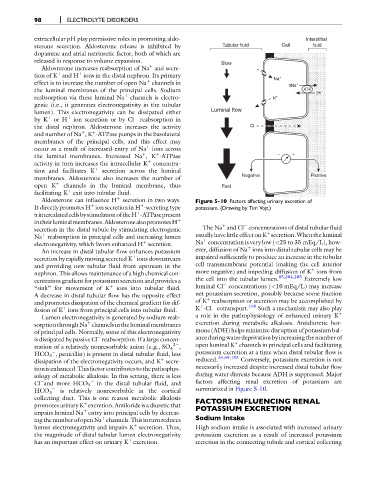
Aldosterone can influence H secretion in two ways. Figure 5-10 Factors affecting urinary excretion of
þ
It directly promotes H ion secretion in H -secreting type potassium. (Drawing by Tim Vojt.)
þ
þ
aintercalatedcellsbystimulationoftheH -ATPasepresent
þ
intheirluminalmembranes.AldosteronealsopromotesH þ
þ
secretion in the distal tubule by stimulating electrogenic The Na and Cl concentrations of distal tubular fluid
þ
þ
Na reabsorption in principal cells and increasing lumen usually have little effect onK secretion.Whenthe luminal
þ
þ
electronegativity, which favors enhanced H secretion. Na concentration is very low (<25 to 35 mEq/L), how-
þ
An increase in distal tubular flow enhances potassium ever, diffusion of Na ions into distal tubular cells may be
secretion by rapidly moving secreted K ions downstream impaired sufficiently to produce an increase in the tubular
þ
and providing new tubular fluid from upstream in the cell transmembrane potential (making the cell interior
þ
nephron. This allows maintenance of a high chemical con- more negative) and impeding diffusion of K ions from
85,204,205
centration gradient for potassium secretion and provides a the cell into the tubular lumen. Extremely low
“sink” for movement of K þ ions into tubular fluid. luminal Cl concentrations (<10 mEq/L) may increase
A decrease in distal tubular flow has the opposite effect net potassium secretion, possibly because some fraction
þ
and promotes dissipation of the chemical gradient for dif- of K reabsorption or secretion may be accomplished by
198
þ
þ
fusion of K ions from principal cells into tubular fluid. K -Cl cotransport. Such a mechanism may also play
þ
Lumen electronegativity is generated by sodium reab- a role in the pathophysiology of enhanced urinary K
þ
sorption through Na channels in the luminal membranes excretion during metabolic alkalosis. Antidiuretic hor-
of principal cells. Normally, some of this electronegativity mone (ADH) helps minimize disruption of potassium bal-
is dissipated by passive Cl reabsorption. If a large concen- ance during water deprivation by increasing the number of
þ
2 open luminal K channels in principal cells and facilitating
tration of a relatively nonresorbable anion (e.g., SO 4 ,
HCO 3 , penicillin) is present in distal tubular fluid, less potassium excretion at a time when distal tubular flow is
36,69,183
þ
dissipation of the electronegativity occurs, and K secre- reduced. Conversely, potassium excretion is not
tion isenhanced.Thisfactor contributestothepathophys- necessarily increased despite increased distal tubular flow
iology of metabolic alkalosis. In this setting, there is less during water diuresis because ADH is suppressed. Major
factors affecting renal excretion of potassium are
Cl and more HCO 3 in the distal tubular fluid, and
summarized in Figure 5-10.
HCO 3 is relatively nonresorbable in the cortical
collecting duct. This is one reason metabolic alkalosis FACTORS INFLUENCING RENAL
promotesurinaryK excretion.Amilorideisadiuretic that POTASSIUM EXCRETION
þ
impairs luminal Na entry into principal cells by decreas-
þ
þ
ingthenumberofopenNa channels.Thisinturnreduces Sodium Intake
þ
lumen electronegativity and impairs K secretion. Thus, High sodium intake is associated with increased urinary
the magnitude of distal tubular lumen electronegativity potassium excretion as a result of increased potassium
þ
has an important effect on urinary K excretion. secretion in the connecting tubule and cortical collecting