
Page 107 - Fluid, Electrolyte, and Acid-Base Disorders in Small Animal Practice
P. 107
Disorders of Potassium: Hypokalemia and Hyperkalemia 97
þ
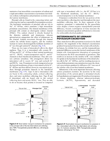
maintains a low intracellular concentration of sodium and with type a intercalated cells (i.e., the H -ATPase is in
a high intracellular concentration of potassium that facili- the basolateral membrane, and the Cl - HCO 3
tate sodium reabsorption and potassium secretion across countertransporter is in the luminal membrane).
the luminal membranes. Potassium is reabsorbed from the last portion of the
Principal cells are found in the connecting tubule and outer medullary collecting duct and throughout the inner
collecting duct and are responsible for potassium secretion. medullary collecting duct. In these segments of the
The basolateral membranes of principal cells are rich in nephron, potassium is reabsorbed by the paracellular
þ
Na ,K -ATPase, which maintains a high intracellular route despite a lumen-negative transepithelial potential
þ
potassium concentration. The luminal membranes of the difference, because reabsorption of water increases the
principal cells contain an electrogenic sodium channel chemical concentration gradient sufficiently to overcome
(ENaC). This sodium channel is directly blocked by the unfavorable electrical gradient.
the diuretics amiloride and triamterene, whereas
spironolactone antagonizes the effect of aldosterone on DETERMINANTS OF URINARY
the channel. Sodium movement through this channel POTASSIUM EXCRETION
renders the tubular lumen negative, and the resultant Three main factors affect potassium secretion in the distal
increase in lumen electronegativity facilitates secretion of nephron: the magnitude of the chemical concentration
þ
þ
K ions through luminal K channels (Fig. 5-8). gradientforpotassium betweenthetubularcellsandtubu-
There are two types of intercalated cells in the distal lar lumen, the tubular flow rate, and the transmembrane
þ
nephron. Type A or a intercalated cells contain H - potential difference across the luminal membranes of the
þ
þ
ATPase and H ,K -ATPase in their luminal membranes tubular cells. Gastrointestinal absorption of a potassium
and Cl - HCO 3 countertransporters and Cl and K þ load increases the ECF concentration of potassium. This
þ
channels in their basolateral membranes. They also con- results in an increase in the number of K ions available
tain carbonic anhydrase. This arrangement allows the for uptake at the basolateral membranes of the distal tubu-
intercalated cell to secrete H þ ions and reabsorb K þ lar cells by Na ,K -ATPase, and the resulting increase in
þ
þ
and HCO 3 ions. Potassium is actively transported across intracellular potassium concentration increases the chem-
the luminal membranes of type a intercalated cells by H , ical concentration gradient for diffusion of K ions out of
þ
þ
þ
K -ATPase and then diffuses down its concentration gra- the tubular cells across their luminal membranes.
dient through potassium channels in the basolateral Aldosterone is the most important hormone affecting
membranes (Fig. 5-9). Type A and a intercalated cells urinary potassium excretion. Its secretion by the zona
are found in the connecting tubule, cortical collecting glomerulosa of the adrenal gland is stimulated directly
duct, and outer medullary collecting duct. Type B and by hyperkalemia and angiotensin II (produced in response
b intercalated cells are found only in the cortical to volume depletion), whereas adrenocorticotropic
collecting ducts and secrete HCO 3 ions. They are able hormone (ACTH), hyponatremia, and decreased
to do so because their polarity is reversed as compared
a Intercalated Interstitial
Interstitial Tubular fluid cell fluid
Tubular fluid Principal Cell fluid
3Na +
Na + ATP
H + 2K +
3Na + ATP K +
Inhibited by ATP + + –
+
K -sparing diuretics 2K TEPD H + OH HOH
Lumen negative CA +CO 2
K + ATP H + HCO 3 –
K + K + Cl –
TEPD K + Cl –
Lumen negative
K +
Figure 5-8 Renal tubular transport mechanisms for potassium Figure 5-9 Renal tubular transport mechanisms for potassium in
in the principal cells of the late distal tubule and collecting the a intercalated cells of the late distal tubule and collecting
duct. TEPD, Transepithelial potential difference. (Drawing by duct. CA, Carbonic anhydrase; TEPD, Transepithelial potential
Tim Vojt.) difference. (Drawing by Tim Vojt.)