
Page 283 - Fluid, Electrolyte, and Acid-Base Disorders in Small Animal Practice
P. 283
274 ACID-BASE DISORDERS
CLINICAL FEATURES OF METABOLIC usually chronic vomiting of stomach contents. Thus, an
ALKALOSIS accurate history is the key to suspecting the diagnosis.
Metabolic alkalosis also can be suspected from the results
The clinical features of dogs and cats with metabolic alka-
of routine serum biochemical tests. Blood gas analysis
losis are usually those of the underlying disease process.
should be performed if decreased serum chloride and
Neurologic signs have been reported in human patients
with severe metabolic alkalosis and include agitation, potassium concentrations are observed and total CO 2
disorientation, stupor, and coma. 103 Muscle twitching content is increased. Blood gas analysis allows the clini-
cian to determine whether primary metabolic alkalosis
and seizures may occur but have been observed rarely
is present and whether the magnitude of respiratory com-
in dogs with severe metabolic alkalosis.
Clinical signs also may result from the accompanying pensation is as predicted (see earlier). The concentration
potassium depletion. Signs of potassium depletion of unmeasured anions (i.e., anion gap) in metabolic alka-
include muscle weakness of varying severity, cardiac losis may increase because of loss of hydrogen ions from
arrhythmias, alterations in renal function (e.g., defective nonbicarbonate buffers. The increased anion gap is pri-
marily caused by increased numbers of negative charges
concentrating ability), and gastrointestinal motility
on proteins and partially the result of the increase in
disturbances (e.g., ileus). These complications are
plasma protein concentration that occurs as a conse-
discussed in Chapter 5. 4
quence of ECFV depletion.
Muscle twitching may occur as a result of decreased
Urine pH is low during the maintenance phase of met-
serum ionized calcium concentration because alkalosis
þ
abolic alkalosis because of enhanced distal Na -H þ
increases the number of negative charges on proteins,
exchange and reabsorption of all filtered HCO 3 . How-
allowing more calcium ions to be bound (Fig. 10-8).
ever, urine pH is alkaline during development of and
Serum ionized calcium concentration decreases and
may account for neuromuscular irritability by rendering recovery from metabolic alkalosis. Thus, urinary pH is
the threshold potential of cells more negative (i.e., bring- of little diagnostic significance in metabolic alkalosis.
ing the resting potential closer to the threshold potential)
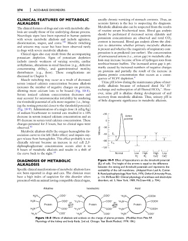
(Fig. 10-9). Administration of a single dose (4 mEq/kg)
of sodium bicarbonate to normal cats resulted in a 10% +30
decrease in serum ionized calcium concentration and an Action
potential
8% decrease in serum total calcium concentration. These 0
changes persisted for 3 hours, but no clinical signs were
observed. 41 –30
Metabolic alkalosis shifts the oxygen-hemoglobin dis- Millivolts
sociation curve to the left (Bohr effect) and impairs oxy- –60 E t
gen release from hemoglobin. This effect probably is not
clinically relevant because an increase in red cell 2,3- –90 E m
diphosphoglycerate concentration occurs after 6 to
–120
8 hours of metabolic alkalosis and results in a shift of
the curve back to the right. 18 Normal Low High High Low
+ + 2+ 2+
K e K e Ca e Ca e
DIAGNOSIS OF METABOLIC Figure 10-9 Effect of hypocalcemia on the threshold potential
ALKALOSIS (E t ) of cells. The height of the arrows is equal to the difference
between the resting and threshold potentials and represents the
Specific clinical manifestations of metabolic alkalosis have excitability of the cell membrane. (Adapted from Leaf A, Cotran
not been reported in dogs and cats. The clinician must R: Renal pathophysiology, New York, 1976, Oxford University Press,
have a high index of suspicion for this disorder when p. 116. IN Rose BD: Clinical physiology of acid-base and electrolyte
presented with an animal having compatible clinical signs, disorders, ed. 3, New York, 1989, McGraw-Hill, p. 704.)
Alkaline Isoelectric Acid
H N COO – + H N COO – + H N COOH
2
3
3
H N COO – + H N COO – + H N COOH
2
H + 3 H + 3
Protein Protein Protein
OH – OH –
N – + – +
H 2 COO H N COO H N COOH
3
3
+ H N COO – + H N COO – + H N COO –
3
3
3
Figure 10-8 Effects of alkalosis and acidosis on the charge of plasma proteins. (Modified from Pitts RF.
Physiology of the kidney and body fluids, 2nd ed. Chicago: Year Book Medical, 1974: 186.)