Page 255 - Feline Cardiology
P. 255
262  Section G: Congestive Heart Failure


                                  Mitral        Start            coronary  arteries  by  concentric  hypertrophy  and/or
                           Aortic  Valve  End of  Atrial  Mitral  increased  left  ventricular  filling  pressure  (Shyu  et  al.
                           Valve  Opening  Rapid  Systole  Valve  2002; Kyriakidis et al. 1997; Cannon et al. 1985). ATP
                           Closure      Ventricular  Closure
                                         Filling                 hydrolysis is required for several stages of the relaxation
                                                                 process,  including  dissociation  of  actin  and  myosin,
                         QRS   T                   QRS   T
                    ECG                       P                  calcium  dissociation  from  troponin  C,  and  sequestra-
                       150                                       tion of calcium by SERCA into the sarcoplasmic reticu-
                                                                 lum. Ischemia leads to greater cytosolic ADP and less
              LV Pressure  100                                   ATP, which prolongs relaxation (as well as impairs sys-
               (mmHg)                                            tolic  function).  In  summary,  impaired  relaxation  is
                        50
                                                                 caused  by  altered  calcium  handling  due  to  abnormal
                        0                                        SERCA activity or level, enhanced myofilament sensitiv-
                                                                 ity  to  calcium,  or  myocardial  ischemia.  Impaired  or
                       10                                        delayed  relaxation  not  only  negatively  impacts  early
               LV Volume  5                                      diastole, but increases myocardial stiffness through the
                 (ml)   0                                        continuing interaction of contractile elements and per-
                                                                 sistent development of myocardial tension (Grossman
      Congestive Heart Failure  LV dv/dt  –400 0                 ventricular mass and myocardial composition. Stiffness
                                                                 and Barry 1980).
                                                                   Ventricular  compliance  is  determined  by  extent  of
                      400
                                                                 is the inverse of compliance. Myocardial determinants
                (ml/sec)
                                                                 of stiffness include changes in the extracellular matrix
                                                 Atrial
                                    Rapid Diastasis
                         Isovolumic
                                                                 (titin) and myofilaments. The main component of the
                                  Ventricular
                         Relaxation
                                                                 cardiac extracellular matrix is collagen (Types I and III).
                                    Filling     Systole          composition or isoform switches in cytoskeletal proteins
                                                                 Type I collagen has the tensile strength of steel and is
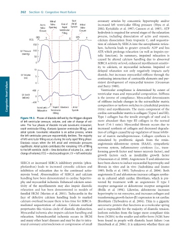
              Figure 19.3.  Phases	of	diastole	defined	by	the	Wiggers	diagram
              of	left	ventricular	pressure,	volume,	and	rate	of	change	of	vol-  more  abundant  than  type  III  collagen  in  the  normal
              ume.	The	four	phases	of	diastole	include	isovolumic	relaxation,	  heart  (7.4 : 1  ratio).  Myocardial  fibrosis  occurs  due  to
              rapid	ventricular	filling,	diastasis	(passive	ventricular	filling),	and	  increased synthesis of collagen and decreased degrada-
              atrial	systole.	Isovolumic	relaxation	is	an	active	process,	where	  tion of collagen caused by up-regulation of tissue inhibi-
              the	left	ventricular	pressure	exponentially	declines.	The	majority	  tor  of  matrix  metalloproteinase.  Collagen  synthesis  is
              of	left	ventricular	filling	occurs	during	the	early	rapid	filling	phase.	  stimulated  by  profibrotic  signals  from  the  renin-
              Diastasis	 occurs	 when	 the	 left	 atrial	 and	 ventricular	 pressures	  angiotensin-aldosterone  system  (RAAS),  sympathetic
              equilibrate.	Atrial	systole	contributes	the	remaining	10%	of	filling	  nervous  system,  inflammatory  cytokines  (i.e.,  trans-
              to	the	left	ventricle.	dv/dt	=	time	derivative	of	volume	(i.e.,	rate	of	  forming growth factor and tumor necrosis factor), and
              change	of	volume);	ECG	=	electrocardiogram	;	LV	=	left	ventricular.
                                                                 growth  factors  such  as  insulinlike  growth  factor
                                                                 (Ouzounian et al. 2008). Angiotensin II and aldosterone
              SERCA or increased SERCA inhibitory protein (phos-  have been shown to induce myocardial hypertrophy and
              pholamban)  leads  to  increased  cytosolic  calcium  and   fibrosis  in  vitro  and  in  vivo  (Sadoshima  and  Izumo
              inhibition  of  relaxation  due  to  the  continued  actin-  1993;  Brilla  et  al.  1993;  Tsybouleva  et  al.  2004).  Both
              myosin  bond.  Abnormalities  of  SERCA  and  calcium   angiotensin II and aldosterone increase collagen synthe-
              handling have been demonstrated in cardiac hypertro-  sis  in  cultured  adult  cardiac  fibroblasts,  which  is  pre-
              phy and myocardial ischemia. Enhanced calcium sensi-  vented  by  treatment  with  an  angiotensin  II  type  I
              tivity  of  the  myofilaments  may  also  impair  diastolic   receptor antagonist or aldosterone receptor antagonist
              relaxation  and  has  been  demonstrated  in  models  of   (Brilla  et  al.  1994).  Likewise,  aldosterone  increases
              familial HCM (Marian et al. 2001). Tachycardia in the   hypertrophy in rat myocytes, and increases collagen and
              face  of  defective  calcium  handling  leads  to  marked   transforming growth factor–β1 expression in rat cardiac
              calcium overload because there is less time for SERCA-  fibroblasts (Tsybouleva et al. 2004). Titin is a gigantic
              mediated  sequestration  of  calcium.  Calcium  overload   sarcomeric protein that functions as a molecular spring
              perpetuates this vicious circle of diastolic dysfunction.   and is responsible for the majority of diastolic tension.
              Myocardial ischemia also impairs calcium handling and   Isoform switches from the larger more compliant titin
              relaxation.  Subendocardial  ischemia  occurs  in  HCM   form (N2BA) to the smaller and stiffer form (N2B) have
              and many other heart diseases and may be due to intra-  been found in people with diastolic heart failure (van
              mural coronary arteriosclerosis or compression of small   Heerebeek et al. 2006). It is unknown whether there are

