Page 256 - Feline Cardiology
P. 256
Chapter 19: Congestive Heart Failure  263


                       End                                       cardiomyopathy)  or  secondary.  Idiopathic  DCM  is  an
                       systole                 Normal            inherent  defect  within  the  myocardium  that  leads  to
                 120                  c        Diastolic dysfunction  myocardial failure. Causative defects may occur within
                             d
                                                                 various regions of the cardiomyocyte, including the con-
                                                                 tractile  sarcomeric  proteins  (affecting  actin-myosin
                LV Pressure (mm Hg)                              production),  cytoskeleton  (disrupting  the  intracellular
                                                                 cross-bridge cycling), mitochondria (impairing energy
                  80
                                                                 and intercellular scaffold and impairing force transmis-
                                                                 sion), nuclear envelope, and transcriptional coactivator
                  40
                             End  b’                             proteins (Maron et al. 2006). Secondary causes of myo-
                             diastole                            cardial failure include nutritional causes (taurine or car-
                  25                                       CHF   nitine  deficiency),  and  diseases  that  lead  to  chronic
                         a’           b
                            a                                    pressure or volume overload of the ventricle (e.g., severe
                                                                 aortic stenosis or ventricular septal defect, respectively),
                             1.5      5
                                                                 and end-stages of other cardiomyopathies (e.g., HCM).
                                   LV Volume (ml)
                                                                 In cats, the only recognized cause of primary myocardial
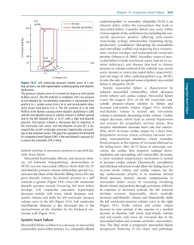
              Figure 19.4.  Left	ventricular	pressure	volume	curve	of	a	nor-  failure is idiopathic DCM.
              mal	cat	and	a	cat	with	hypertrophic	cardiomyopathy	and	diastolic	  Systolic  myocardial  failure  is  characterized  by
              dysfunction.                                       reduced  myocardial  contractility,  which  decreases
              The	pressure	volume	curve	of	a	normal	cat	begins	at	end-systole	  stroke  volume  and  cardiac  output.  Myocardial  failure
              (a	[blue	curve]).	The	left	ventricle	is	compliant,	with	low	pressure	                                     Congestive Heart Failure
              at	end-diastole	(b).	Isovolumetric	contraction	is	represented	from	  causes  the  tangent  (Ees)  of  the  left  ventricular  end-
              points	b	to	c,	systole	occurs	from	c	to	d,	and	isovolumetric	relax-  systolic  pressure-volume  relation  to  flatten  and
              ation	occurs	from	points	d	to	a.	The	left	ventricle	of	a	cat	with	  increases  end-systolic  volume  (Figure  19.5).  Initially,
              HCM	or	other	disease	causing	severe	diastolic	dysfunction	is	stiff,	  end-diastolic  volume  is  normal  and  end-systolic
              and	the	end-diastolic	pressure	volume	relation	is	shifted	upward	  volume is increased, decreasing stroke volume. Cardiac
              and	to	the	left	(dashed	line:	a’	to	b’)	with	a	high	end-diastolic	  output  decreases,  which  leads  to  arterial  hypotension
              pressure.	End-systolic	volume	is	decreased	due	to	reduction	in	  and  activates  the  sympathetic  nervous  system.  Beta
              left	ventricular	wall	stress,	and	end-diastolic	volume	(b’)	is	de-  receptor  stimulation  increases  heart  rate  and  contrac-
              creased	due	to	left	ventricular	concentric	hypertrophy	encroach-  tility, which increases cardiac output for a short time.
              ing	on	the	chamber	lumen.	The	grey	line	represents	the	threshold	  Sympathetic  nervous  system  activation  increases  arte-
              for	congestive	heart	failure	(CHF);	if	the	end-diastolic	pressure	(b’)	  riolar  vasoconstrictor  mediators,  which  normalize
              is	above	this	threshold,	CHF	is	likely.
                                                                 blood pressure, at the expense of increased afterload on
                                                                 the failing heart. After 24–72 hours of adrenergic acti-
              isoform switches in sarcomeric proteins in cats with dia-  vation,  the  cardiac  beta  receptors  undergo  down-
              stolic heart failure.                              regulation and uncoupling, and contractility decreases.
                 Myocardial hypertrophy, fibrosis, and myocyte disar-  A more sustained compensatory mechanism is needed
              ray  (all  hallmark  histopathologic  abnormalities  in   to  increase  cardiac  output.  Theoretically,  vasodilation
              HCM) increase myocardial stiffness, which shifts the LV   and afterload reduction would allow the failing heart to
              end-diastolic pressure volume relationship upward (i.e.,   increase  stroke  volume  and  cardiac  output,  but  the
              increases the slope of the diastolic filling curve). For any   top  cardiovascular  priority  is  to  maintain  normal
              given diastolic volume, the diastolic pressure in a stiff   blood  pressure.  Instead,  chronic  compensation  in
              ventricle is greater (Figure 19.4). Once left ventricular   myocardial  failure  occurs  by  increasing  circulating
              diastolic  pressure  exceeds  25 mm Hg,  left  heart  failure   blood volume and preload through activation of RAAS.
              develops.  Left  ventricular  concentric  hypertrophy   In  response  to  increased  preload,  the  left  ventricle
              decreases  systolic  wall  stress,  which  decreases  end-  develops  eccentric  hypertrophy,  which  markedly
              systolic volume and shifts the left ventricular pressure   increases  end-diastolic  volume  and  dramatically  shifts
              volume curve to the left (Figure 19.4). Left ventricular   the left ventricular pressure volume curve to the right
              end-diastolic  diameter  is  also  decreased  due  to  the   (Figure  19.5).  Stroke  volume  and  cardiac  output
              encroachment  of  the  chamber  by  the  thickened  ven-  increase  to  near  normal  at  the  expense  of  a  marked
              tricular wall (Figure 19.4).                       increase  in  diastolic  wall  stress.  End-systolic  volume
                                                                 and  end-systolic  wall  stress  are  increased  due  to  the
              Systolic heart failure                             myocardial failure and systemic arteriolar vasoconstric-
              Myocardial failure is defined as a decrease in myocardial   tion.  The  final  result  is  progressive  myocardial  failure
              contractility and is either primary (i.e., idiopathic dilated   (progressive  flattening  of  Ees  slope)  and  pathologic

