Page 260 - Feline Cardiology
P. 260
Chapter 19: Congestive Heart Failure  267


              has occurred, often there is pinkish fluid secondary to
              capillary  hemorrhage.  Microscopically,  edema  fluid
              appears  acidophilic  or  faintly  granular,  and  fills  the
              alveoli, interstitium, and lymphatics (Liu and Fox 1999).
              Lymphatics and pulmonary capillaries appear engorged
              and tortuous. Alveolar macrophages containing eryth-
              rocytes  or  hemosiderin  (i.e.,  heart  failure  cells)  are
              present throughout the lungs and increase in number
              with  chronicity  of  heart  failure  (Liu  and  Fox  1999).
              There  are  often  regions  of  red,  well-demarcated,  firm
              tissue  in  the  lung  periphery,  consisting  of  pulmonary
              hemorrhage  and  infarction  (SK  Liu  1970).  Muscular
              hypertrophy  of  small  pulmonary  arteries  and  fibrous
              thickening of pulmonary capillary walls is seen in cats
              with pulmonary hypertension (uncommon).
                 Pleural effusion occurs secondary to left or right heart   A
              failure. Grossly, it may appear clear to tan, or chylous.
              The mechanism of true chylous effusion occurring sec-
              ondary to isolated left-sided cardiac disease is unknown.
              True  chylous  effusion  occurring  secondary  to  right-
              sided heart disease may occur due to increased hydro-                                                     Congestive Heart Failure
              static pressures in the thoracic duct, since the thoracic
              duct empties into the cranial vena cava which has ele-
              vated hydrostatic pressure. The pleura typically appear
              thickened in cases of chronic pleural effusion, and lung
              lobe  edges  may  be  fibrotic.  Lung  lobe  collapse  is  also
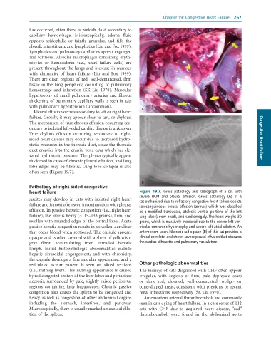
              often seen (Figure 19.7).


                                                                  B
              Pathology of right-sided congestive
              heart failure                                      Figure  19.7.  Gross	 pathology	 and	 radiograph	 of	 a	 cat	 with
                                                                 severe	 HCM	 and	 pleural	 effusion.	 Gross	 pathology	 (A)	 of	 a
              Ascites  may  develop  in  cats  with  isolated  right  heart
              failure and is most often seen in conjunction with pleural   cat	euthanized	due	to	refractory	congestive	heart	failure	depicts
                                                                 serosanguineous	pleural	effusion	(arrows)	which	was	classified
              effusion. In passive hepatic congestion (i.e., right heart   as	 a	 modified	 transudate,	 atelectic	 ventral	 portions	 of	 the	 left
              failure), the liver is heavy (∼115–133 grams), firm, and   lung	lobe	(arrow	head),	and	cardiomegaly.	The	heart	weighs	30
              swollen with rounded edges of the central lobes. Acute   grams,	which	is	massively	increased	due	to	the	severe	left	ven-
              passive hepatic congestion results in a swollen, dark liver   tricular	concentric	hypertrophy	and	severe	left	atrial	dilation.	An
              that oozes blood when sectioned. The capsule appears   antemortem	lateral	thoracic	radiograph	(B)	of	this	cat	provides	a
              opaque and is often covered with a sheet of yellowish-  clinical	correlate,	and	shows	severe	pleural	effusion	that	obscures
              gray  fibrin  accumulating  from  extruded  hepatic    the	cardiac	silhouette	and	pulmonary	vasculature.
              lymph.  Initial  histopathologic  abnormalities  include
              hepatic  sinusoidal  engorgement,  and  with  chronicity,
              the capsule develops a fine nodular appearance, and a
              reticulated  acinar  pattern  is  seen  on  sliced  sections     Other pathologic abnormalities
              (i.e., nutmeg liver). This nutmeg appearance is caused   The kidneys of cats diagnosed with CHF often appear
              by red congested centers of the liver lobes and periacinar   irregular,  with  regions  of  firm,  pale  depressed  scars
              necrosis, surrounded by pale, slightly raised periportal   or  dark  red,  elevated,  well-demarcated,  wedge-  or
              regions  containing  fatty  hepatocytes.  Chronic  passive   cone-shaped  areas,  consistent  with  previous  or  recent
              congestion also causes the spleen to be congested and   renal infarctions, respectively (SK Liu 1970).
              heavy, as well as congestion of other abdominal organs   Antemortem arterial thromboemboli are commonly
              including  the  stomach,  intestines,  and  pancreas.   seen in cats dying of heart failure. In a case series of 112
              Microscopically, there is usually marked sinusoidal dila-  cats  with  CHF  due  to  acquired  heart  disease,  “red”
              tion of the spleen.                                thromboemboli  were  found  in  the  abdominal  aorta

