Page 264 - Feline Cardiology
P. 264
Chapter 19: Congestive Heart Failure  271


              cyanosis and should be quickly triaged for emergency
              care.  Muffled  lung  and  heart  sounds  are  commonly
              present  with  significant  pleural  effusion,  and  often
              patients have shallow, rapid respirations. Palpation of a
              noncompliant cranioventral thorax may raise the suspi-
              cion of a cranial mediastinal mass as the cause of pleural
              effusion. Adventitious  lung  sounds  may  be  present  in
              cats with pulmonary edema and may overshadow detec-
              tion of a murmur.
                 Abnormal cardiac auscultation including a murmur,
              gallop  heart  sound,  or  arrhythmia  is  variably  present.
              Only  20%  of  cats  dying  of  heart  failure  in  one  study
              had an abnormal cardiac auscultation (Liu et al. 1970).
              Factors involved with such a low occurrence of abnor-
              mal  cardiac  auscultation  may  include  muffled  heart
              sounds from pleural effusion, adventitious lung sounds
              masking a murmur, or at the time of that publication
              in the 1960s the high prevalence of dilated cardiomy-
              opathy, which less commonly causes a murmur. A sys-
              tolic murmur may be more commonly present in cats
              with HCM (64–72%) than in cats with restrictive car-        A                                             Congestive Heart Failure
              diomyopathy  (36%)  or  dilated  cardiomyopathy  (9%)
              (Rush  et  al.  2002;  Ferasin  et  al.  2003).  Likewise,  the
              murmur  intensity  is  typically  loudest  in  cats  with
              hypertrophic obstructive cardiomyopathy compared to
              other cardiac diseases. A gallop heart sound (i.e., extra
              S 3   or  S 4   heart  sound)  is  more  common  in  cats  with
              DCM (45%) compared to HCM (12–33%) and RCM
              (23%) (see Chapter 1) (Rush et al. 2002; Ferasin et al.
              2003). Tachycardia is commonly present and is a result
              of sympathetic activation (i.e., sinus tachycardia) or a
              tachyarrhythmia  (i.e.,  supraventricular  tachycardia,
              ventricular tachycardia, or atrial fibrillation).
                 Lethargy, weakness, collapse, or poor femoral arterial
              pulse  quality  may  be  present  in  cats  with  low  output
              heart failure, which is more common in cats with DCM
              than other cardiac diseases. Slow capillary refill time and
              pale mucous membranes occur when there is low cardiac
              output  and  peripheral  hypoperfusion.  Absence  of   B
              femoral  arterial  pulses,  cold  extremeties,  and  cyanotic
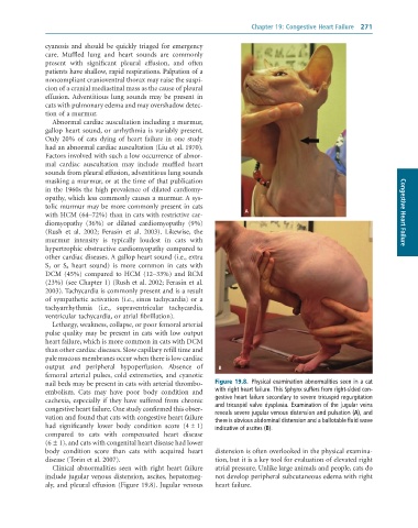
              nail beds may be present in cats with arterial thrombo-  Figure 19.8.  Physical	examination	abnormalities	seen	in	a	cat
              embolism.  Cats  may  have  poor  body  condition  and   with	right	heart	failure.	This	Sphynx	suffers	from	right-sided	con-
              cachexia, especially if they have suffered from chronic   gestive	heart	failure	secondary	to	severe	tricuspid	regurgitation
              congestive heart failure. One study confirmed this obser-  and	tricuspid	valve	dysplasia.	Examination	of	the	jugular	veins
              vation and found that cats with congestive heart failure   reveals	severe	jugular	venous	distension	and	pulsation	(A),	and
                                                                 there	is	obvious	abdominal	distension	and	a	ballotable	fluid	wave
              had  significantly  lower  body  condition  score  (4 ± 1)   indicative	of	ascites	(B).
              compared  to  cats  with  compensated  heart  disease
              (6 ± 1), and cats with congenital heart disease had lower
              body  condition  score  than  cats  with  acquired  heart   distension is often overlooked in the physical examina-
              disease (Torin et al. 2007).                       tion, but it is a key tool for evaluation of elevated right
                 Clinical  abnormalities  seen  with  right  heart  failure   atrial pressure. Unlike large animals and people, cats do
              include jugular venous distension, ascites, hepatomeg-  not develop peripheral subcutaneous edema with right
              aly, and pleural effusion (Figure 19.8). Jugular venous   heart failure.

