Page 267 - Feline Cardiology
P. 267
274  Section G: Congestive Heart Failure


                      A


      Congestive Heart Failure


                      C
                                                                      B
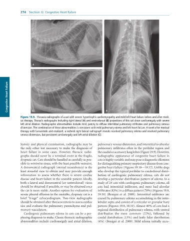
              Figure 19.9.  Thoracic	radiographs	of	a	cat	with	severe	hypertrophic	cardiomyopathy	and	mild	left	heart	failure	before	and	after	medi-
              cal	therapy.	Thoracic	radiographs	including	right	lateral	(A)	and	ventrodorsal	(B)	projections	of	this	cat	show	cardiomegaly	with	severe
              left	atrial	dilation.	Radiographic	abnormalities	include	mild,	patchy	to	diffuse	interstitial	pulmonary	infiltrates	and	pulmonary	venous
              distension.	The	combination	of	these	abnormalities	is	consistent	with	mild	pulmonary	edema	and	left	heart	failure.	A	week	after	medical
              therapy	with	furosemide	and	enalapril,	a	recheck	right	lateral	radiograph	reveals	resolved	pulmonary	edema	and	resolved	pulmonary
              venous	distension,	but	persistent	cardiomegaly	and	left	atrial	dilation	(C).


              history and physical examination, radiographs may be   pulmonary venous distension, and interstitial to alveolar
              the only other test necessary to make the diagnosis of   pulmonary infiltrates often in the perihilar region and
              heart  failure  in  some  cases.  However,  thoracic  radio-  the caudal or accessory lung lobes (Figure 19.9). However,
              graphs should never be a terminal event in the fragile,   radiographic appearance of congestive heart failure in
              dyspneic cat. Cats should be handled as carefully as pos-  cats is highly variable and may pose a diagnostic dilemma
              sible to minimize stress, with the least possible restraint.   for distinguishing primary respiratory disease from con-
              A dorsoventral radiograph (sternal recumbency) is the   gestive heart failure (Figures 19.10—19.12). Unlike dogs
              least stressful view to obtain and may provide enough   who develop the typical perihilar to caudodorsal distri-
              information  to  assess  whether  there  is  severe  cardiac   bution  of  cardiogenic  pulmonary  edema,  cats  do  not
              disease and heart failure in the unstable patient. Ideally,   develop a particular distribution pattern of edema. In a
              both a lateral and dorsoventral (or ventrodorsal) views   study of 23 cats with cardiogenic pulmonary edema, all
              should be obtained if possible, or may be obtained once   cats  had  interstitial  infiltrates,  and  most  had  alveolar
              the cat is more stable. Another option for evaluation of   infiltrates (83%) in a diffuse pattern (78%) (Figures 19.9,
              severe pleural effusion in the unstable, dyspneic cat is a   19.10)  (Benigni  et  al.  2009).  Interstitial  infiltrates  are
              brief  “triage”  echocardiogram.  Two-view  radiographs   caused by pulmonary edema accumulating in the inter-
              should be obtained after thoracocentesis to assess cardiac   lobular septa and consist of a reticular or granular hazy
              size and evaluate the pulmonary parenchyma and pul-  pattern (Figures 19.9, 19.10). Almost 40% of cats had a
              monary vasculature.                                regional distribution of pulmonary edema, with ventral
                 Cardiogenic pulmonary edema in cats can be a per-  distribution  the  most  common  (22%),  followed  by
              plexing diagnosis to make. Classic thoracic radiographic   caudal distribution (13%) and lastly hilar distribution
              abnormalities include cardiomegaly and atrial dilation,   (4%) (Benigni et al. 2009). Mild edema initially accu-

