Page 268 - Feline Cardiology
P. 268
Chapter 19: Congestive Heart Failure  275


                A                                                 C                                                     Congestive Heart Failure


                            B                                 D
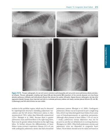
              Figure 19.10.  Thoracic	radiographs	of	a	cat	with	severe	restrictive	cardiomyopathy	and	concurrent	severe	pulmonary	edema	and	pleu-
              ral	effusion.	Thoracic	radiographs	including	right	lateral	(A)	and	dorsoventral	(B)	projections	of	this	severely	dyspneic	cat	show	heavy
              alveolar	pulmonary	infiltrates	and	mild	pleural	effusion	that	obscure	the	cardiac	silhouette.	Repeat	thoracic	radiographs	one	day	after
              aggressive	diuretic	therapy	show	improved	and	mild	to	moderate	pulmonary	edema	and	nearly	resolved	pleural	effusion	(C)	and	(D).
              Cardiomegaly	and	left	atrial	dilation	are	now	visible.


              mulates in the perihilar region, which may be obscured   pulmonary  pattern  (Benigni  et  al.  2009).  Cardiogenic
              by superimposed structures including pulmonary vas-  pulmonary edema was never present in just a single lung
              culature and the left atrium. Pulmonary edema is often   lobe, which may help distinguish heart failure from some
              asymmetrical (78%) rather than bilaterally symmetrical   cases of bronchopneumonia or aspiration pneumonia.
              (22%)  (Benigni  et  al.  2009).  Because  there  is  greater   Although often present in heart failure (71% of cats in
              pulmonary mass and blood flow to the right lung lobes,   1  study),  pulmonary  venous  distension  may  not  be
              there may be greater edema formation in the right lung   present in cats that are dehydrated or receiving diuretics
              lobes.  To  further  obscure  the  differentiation  of  heart   (Benigni et al. 2009). Often both pulmonary arteries and
              failure versus primary respiratory disease, 61% of cats   pulmonary  veins  are  distended  in  congestive  heart
              with cardiogenic pulmonary edema also had a bronchial   failure. Heart failure should never be ruled out based on

