Page 269 - Feline Cardiology
P. 269
276  Section G: Congestive Heart Failure


                   A                                                 C


      Congestive Heart Failure


                                B                               D
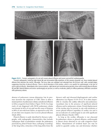
              Figure 19.11.  Thoracic	radiographs	of	a	cat	with	severe	pleural	effusion	and	severe	unclassified	cardiomyopathy.
                 Thoracic	radiographs	including	right	lateral	(A)	and	dorsoventral	(B)	projections	of	this	severely	dyspneic	cat,	show	marked	pleural
              effusion	that	obscures	the	cardiac	silhouette.	The	lung	lobes	are	scalloped	and	retracted	from	the	thoracic	wall,	with	rounded	edges
              suggesting	chronicity	of	pleural	effusion.	There	is	dorsal	deviation	of	the	trachea,	which	is	not	specific	for	cardiomegaly	in	the	face	of
              severe	pleural	effusion.	Immediately	after	thoracocentesis,	right	lateral	and	ventrodorsal	projections	show	mild	residual	pleural	effusion
              (C)	and	(D).	Biatrial	dilation	and	severe	cardiomegaly	are	present,	as	well	as	moderate,	patchy	to	diffuse	pulmonary	infiltrates	consistent
              with	pulmonary	edema.


              absence of pulmonary venous distension, but its pres-  thoracic wall, and obscured diaphragmatic and cardiac
              ence  increases  the  suspicion  of  CHF.  There  is  often  a   silhouettes (see Figures 19.10–19.12). It is often impos-
              mixed pattern of pulmonary edema and pleural effusion   sible to visualize the cardiac silhouette and pulmonary
              in feline congestive heart failure (Figure 19.10). In a large   vasculature  due  to  the  presence  of  significant  pleural
              study of cats diagnosed with HCM, pulmonary edema   effusion and/or pulmonary edema. Dorsal displacement
              was present in 66% of cats and was the cause of dyspnea   of the trachea may be present in cats with moderate or
              in 86% of cats with heart failure, as opposed to only 14%   severe pleural effusion regardless of cardiac size and is
              of cats that were dyspneic due to severe pleural effusion   not an indicator of cardiomegaly in cats with significant
              (Rush 2002).                                       pleural effusion (Snyder 1990).
                 Pleural effusion is easily identified by thoracic radio-  As  long  as  the  cardiac  silhouette  is  not  obscured
              graphs,  with  radiographic  characteristics  that  include:   by pulmonary edema or pleural effusion, cardiomegaly
              radiopaque fluid accumulation outside the pulmonary   is almost always detected in cats with congestive heart
              parenchyma, pleural fissure lines, scalloping (rounding)   failure.  Measurement  of  vertebral  heart  size  may  be
              of the lung lobes, retraction of the lung lobes from the   useful to quantify cardiac size and determine whether

