Page 270 - Feline Cardiology
P. 270
Chapter 19: Congestive Heart Failure  277


                         A


                                                                        B                                               Congestive Heart Failure


                                      C


                         D                                               E
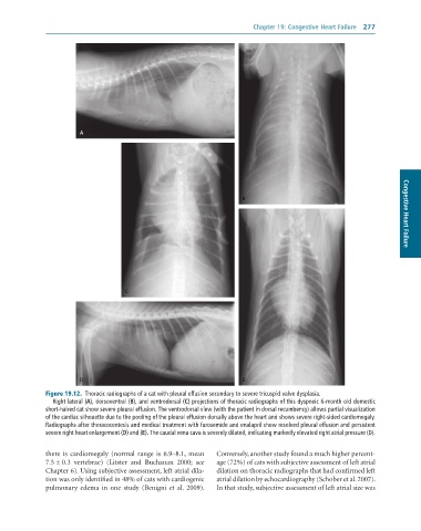
              Figure 19.12.  Thoracic	radiographs	of	a	cat	with	pleural	effusion	secondary	to	severe	tricuspid	valve	dysplasia.
                 Right	lateral	(A),	dorsoventral	(B),	and	ventrodorsal	(C)	projections	of	thoracic	radiographs	of	this	dyspneic	6-month	old	domestic
              short-haired	cat	show	severe	pleural	effusion.	The	ventrodorsal	view	(with	the	patient	in	dorsal	recumbency)	allows	partial	visualization
              of	the	cardiac	silhouette	due	to	the	pooling	of	the	pleural	effusion	dorsally	above	the	heart	and	shows	severe	right-sided	cardiomegaly.
              Radiographs	after	thoracocentesis	and	medical	treatment	with	furosemide	and	enalapril	show	resolved	pleural	effusion	and	persistent
              severe	right	heart	enlargement	(D)	and	(E).	The	caudal	vena	cava	is	severely	dilated,	indicating	markedly	elevated	right	atrial	pressure	(D).


              there  is  cardiomegaly  (normal  range  is  6.9–8.1,  mean   Conversely, another study found a much higher percent-
              7.5 ± 0.3  vertebrae)  (Litster  and  Buchanan  2000;  see   age (72%) of cats with subjective assessment of left atrial
              Chapter 6). Using subjective assessment, left atrial dila-  dilation on thoracic radiographs that had confirmed left
              tion was only identified in 48% of cats with cardiogenic   atrial dilation by echocardiography (Schober et al. 2007).
              pulmonary  edema  in  one  study  (Benigni  et  al.  2009).   In that study, subjective assessment of left atrial size was

