Page 257 - Feline Cardiology
P. 257
264  Section G: Congestive Heart Failure


                                             Normal                                            Normal
                                             Chronic myocardial failure  End                   Patent ductus arteriosus
                     End                     Acute myocardial failure    systole
                     systole
                 120                c                               120    Ees  d                    c’
                        Ees  d                                                      d’    c
                LV Pressure (mm Hg)  80  d’  c’                   LV Pressure (mm Hg)  80


                               End
                  40
                               diastole                                             End
                                    b’                              40              diastole         b’
                  25                b                CHF
                          a  a’                                     25                    b                  CHF
                                                                               a     a’
                           1.5     5      10    15                                                       f
                               LV Volume (ml)                                   1.5      5        10     15
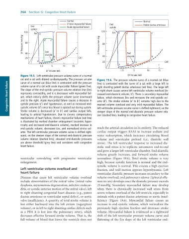
               Figure 19.5.  Left	ventricular	pressure	volume	curve	of	a	normal	     LV Volume (ml)
               cat	and	a	cat	with	dilated	cardiomyopathy.	The	pressure	volume	  Figure 19.6.  The	pressure	volume	curve	of	a	normal	cat	(blue
      Congestive Heart Failure  The	slope	of	the	end-systolic	pressure	volume	relation	line	(Ees)	  to	right	shunt	causes	severe	left	ventricular	volume	overload	(in-
               curve	of	a	normal	cat	(blue	line)	is	contrasted	with	the	pressure
                                                                 line)	is	contrasted	with	the	curve	of	a	cat	with	a	large	left	to
               volume	curve	of	a	cat	with	acute	myocardial	failure	(green	line).
                                                                 right	shunting	patent	ductus	arteriosus	(red	line).	The	large	left
               represents	contractility,	and	it	is	decreased	with	myocardial	fail-
                                                                 creased	end-diastolic	volume,	b′).	There	is	secondary	myocardial
               ure,	which	initially	shifts	the	pressure	volume	curve	downward
                                                                 failure,	which	decreases	Ees	and	increases	the	end-systolic	vol-
               and	to	the	right.	Acute	myocardial	failure	causes	a	decrease	in
                                                                 ume	(d′).	The	stroke	volume	(a′	to	b′)	remains	high	due	to	the
               systolic	pressure	(c′)	and	hypotension,	as	well	as	increased	end-
                                                                 marked	volume	overload	and	only	mild	myocardial	failure.	The
               systolic	volume	(d′)	since	less	blood	is	ejected	out	during	systole.
               Stroke	volume	is	decreased	(a′	to	b′)	and	cardiac	output	falls,
                                                                 steeper	slope	of	the	normal	end-diastolic	pressure	volume	rela-
               leading	 to	 arterial	 hypotension.	 Due	 to	 chronic	 compensatory	  left	ventricular	pressure	volume	curve	is	shifted	rightward,	on	the
                                                                 tion	(dashed	line),	leading	to	congestive	heart	failure.
               mechanisms	of	heart	failure,	chronic	myocardial	failure	(red	line)
               is	illustrated	by	marked	chamber	enlargement	(eccentric	hyper-
               trophy	and	increased	end-diastolic	volume),	marked	increase	in
               end-systolic	volume,	decreased	Ees,	and	normalized	stroke	vol-  reach the arterial circulation in its entirety. The reduced
               ume.	The	left	ventricular	pressure	volume	curve	is	shifted	right-  cardiac  output  triggers  RAAS  to  increase  sodium  and
               ward,	on	the	steeper	slope	of	the	normal	end-diastolic	pressure	  water  reabsorption,  which  increases  circulating  blood
               volume	 relation	 (dashed	 line);	 elevated	 end-diastolic	 pressures	  volume  and  ventricular  preload  (i.e.,  diastolic  wall
               are	above	threshold	(grey	line)	and	consistent	with	congestive	  stress).  The  left  ventricular  response  to  increased  dia-
               heart	failure.                                    stolic wall stress is to replicate sarcomeres end-to-end
                                                                 and grow a larger left ventricular chamber. End-diastolic
                                                                 volume  greatly  increases,  and  forward  stroke  volume
              ventricular  remodeling  with  progressive  ventricular   normalizes  (Figure  19.6).  Total  stroke  volume  is  very
              enlargement.                                       high, because systolic function is normal and the end-
                                                                 systolic volume is normal. This creates a high ejection
              Left ventricular volume overload and               fraction, and wall motion appears hyperdynamic. Left
              heart failure                                      ventricular diastolic pressure increases secondary to the
              Diseases  that  cause  left  ventricular  volume  overload   volume overload, and pulmonary edema (±pleural effu-
              include abnormalities of the mitral valve (mitral valve   sion in cats) develops once the diastolic pressure exceeds
              dysplasia, myxomatous degeneration, infective endocar-  25 mm Hg.  Secondary  myocardial  failure  may  develop
              ditis, or systolic anterior motion of the mitral valve), left   when  there  is  chronically  increased  wall  stress  from
              to right shunting congenital heart diseases (e.g., patent   severe volume overload of the left ventricle, especially in
              ductus arteriosus or ventricular septal defect), or aortic   animals with a patent ductus arteriosus or aortic insuf-
              valve insufficiency. A quantity of total stroke volume is   ficiency  (Figure  19.6).  Myocardial  failure  causes  an
              lost  either  backward  into  the  left  atrium  (regurgitant   increase in end-systolic volume, which normalizes the
              volume), or in left to right shunting cardiac defects such   previously  high  ejection  fraction  and  decreases  stroke
              as  a  PDA  it  is  lost  into  the  pulmonary  artery,  which   volume. Myocardial failure is evidenced as a rightward
              decreases effective forward stroke volume. That is, the   shift of the left ventricular pressure volume curve and
              full volume of blood that leaves the ventricle does not   flattening of the Ees slope of the left ventricular end-

