Page 298 - Feline Cardiology
P. 298
306  Section H: Arterial Thromboembolism


                                                                 contrast (SEC) in cats with cardiomyopathy. In fact, left
                                                                 auricular velocity <0.20 m/s was the only independent
                                                                 variable that predicted SEC. SEC is a possible indicator
                                                                 that  a  cat  is  at  increased  risk  for ATE  (Schober  et  al.
                                                                 2006).
                                                                   Results regarding hypercoagulability in cats with heart
                                                                 disease have been variable; however, a hypercoagulable
                                                                 environment is present in some cats. For example, feline
                                                                 platelets are particularly reactive and responsive to ade-
                                                                 nosine diphosphate (ADP) and other agonists of platelet
                                                                 aggregation. One study showed that platelets from car-
                                                                 diomyopathic cats required less ADP to induce aggrega-
                                                                 tion than did platelets from normal cats (Helinski and
                                                                 Ross 1987), while another demonstrated decreased plate-
                                                                 let responsiveness to ADP, but increased responsiveness
                                                                 to  collagen  (Welles  et  al.  1994). Another  study,  which
                                                                 defined systemic hypercoagulability as two or more labo-
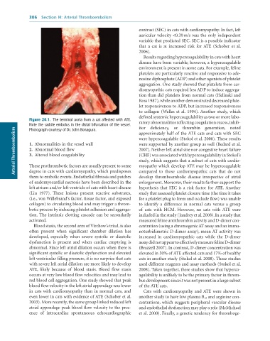
              Figure 20.1.  The	terminal	aorta	from	a	cat	affected	with	ATE.	  ratory abnormalities reflecting coagulation excess, inhib-
              Note	the	saddle	embolus	in	the	distal	bifurcation	of	the	vessel.	  itor  deficiency,  or  thrombin  generation,  noted
        Arterial Thromboembolism  1.  Abnormalities in the vessel wall  were hypercoagulable (Stokol et al. 2008). These results
              Photograph	courtesy	of	Dr.	John	Bonagura.
                                                                 approximately half of the ATE cats and cats with SEC

                                                                 were supported by another group as well (Bedard et al.
                                                                 2007). Neither left atrial size nor congestive heart failure
              2.  Abnormal blood flow
              3.  Altered blood coagulability
                                                                 (CHF) was associated with hypercoagulability in Stokol’s
                                                                 study, which suggests that a subset of cats with cardio-
              These prothrombotic factors are usually present to some
              degree in cats with cardiomyopathy, which predisposes
                                                                 compared  to  those  cardiomyopathic  cats  that  do  not
              them to embolic events. Endothelial fibrosis and patches   myopathy which develop ATE may be hypercoagulable
                                                                 develop  thromboembolic  disease  irrespective  of  atrial
              of endomyocardial necrosis have been described in the   enlargement. Moreover, their results further support the
              left atrium and/or left ventricle of cats with heart disease   hypothesis  that  SEC  is  a  risk  factor  for ATE. Another
              (Liu  1977).  These  lesions  present  reactive  substrates,   study that assessed platelet closure time (the time it takes
              (i.e., von Willebrand’s factor, tissue factor, and exposed   for a platelet plug to form and occlude flow) was unable
              collagen) to circulating blood and may trigger a throm-  to  identify  a  difference  in  normal  cats  versus  a  group
              botic process by inducing platelet adhesion and aggrega-  of  cats  with  HCM.  However,  no  cats  with  ATE  were
              tion. The intrinsic clotting cascade can be secondarily   included in the study (Jandrey et al. 2008). In a study that
              activated.                                         measured feline antithrombin activity and D-dimer con-
                 Blood stasis, the second arm of Virchow’s triad, is also   centration (using a chromogenic AT assay and an immu-
              often  present  when  significant  chamber  dilation  has   noturbidimetric D-dimer assay), mean AT activity was
              developed,  especially  when  severe  systolic  or  diastolic   increased  in  cardiomyopathic  cats  while  the  D-dimer
              dysfunction  is  present  and  when  cardiac  emptying  is   assay did not appear to effectively measure feline D-dimer
              abnormal. Since left atrial dilation occurs when there is   (Brazzell 2007). In contrast, D-dimer concentration was
              significant systolic or diastolic dysfunction and elevated   elevated in 50% of ATE affected cats and 17% of healthy
              left ventricular filling pressure, it is no surprise that cats   cats in another study (Stokol et al. 2008). These studies
              with severe left atrial dilation are more likely to develop   used different reagents and assay methods (Stokol et al.
              ATE,  likely  because  of  blood  stasis.  Blood  flow  stasis   2008). Taken together, these studies show that hyperco-
              occurs at very low blood flow velocities and may lead to   agulability is unlikely to be the primary factor in throm-
              red blood cell aggregation. One study showed that peak   bus development since it was not present in a large subset
              blood flow velocity in the left atrial appendage was lower   of the ATE cats.
              in cats with cardiomyopathy than in normal cats, and   Cats  with  cardiomyopathy  and ATE  were  shown  in
              even lower in cats with evidence of ATE (Schober et al.   another study to have low plasma B 12  and arginine con-
              2003). More recently, the same group linked reduced left   centrations, which suggests peripheral vascular disease
              atrial appendage peak blood flow velocity to the pres-  and endothelial dysfunction may play a role (McMichael
              ence  of  intracardiac  spontaneous  echocardiographic   et al. 2000). Finally, a genetic tendency for thromboge-

