Page 303 - Feline Cardiology
P. 303
Chapter 20: Arterial Thromboembolism  311


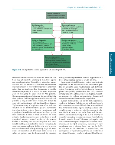
                                                    Physical examination
                                                    Rectal temperature and number
                                                    of limbs affected for prognosis


                                    Initial diagnostics             Emergency treatment
                                    -Thoracic radiographs           -Therapy for congestive heart failure
                                    -ECG if arrhythmias or bradycardia  if indicated by radiographs
                                    -Serum chemistry                (see CHF chapter for details)
                                    -Urinalysis                     -Analgesia therapy


                                    Follow-up diagnostics           Short-term management
                                    -Cardiology work up             -Heart failure medications if indicated
                                    -Thyroid status (depending on age)  -Fluid therapy if indicated
                                    -If no heart disease, search for  -Heparin therapy
                                    underlying cause of thromboembolism  -Nutritional support
                                    (i.e. neoplasia, etc.)          -Rehabilitation therapy


                                                  Long-term management
                                                  -Cardiac medications if indicated
                                                  -Gradually discontinue heparin after
                                                  beginning long-term anticoagulant therapy
                                                  -Rehabilitation therapy                                               Arterial Thromboembolism
                                                  -Wound management if ischemic necrosis

              Figure 20.8.  An	algorithm	for	a	clinical	approach	for	cats	presenting	with	ATE.


              rial vasodilation is often not uniform and flow to muscle   licking or chewing of the toes or hock. Application of a
              beds  may  ultimately  be  unchanged. Also,  these  agents   loose fitting bandage barrier is usually effective.
              can cause hypotension. Their efficacy is therefore uncer-  Appropriate clinical laboratory patient monitoring is
              tain and their use has fallen out of favor. Hypothermia   dependent on the individual’s status. Biochemical pro-
              is a manifestation of poor systemic perfusion and shock   files are useful to assess renal function and electrolyte
              rather than just local blood flow changes due to a saddle   status. Coagulation profiles (activated partial thrombo-
              thrombus; improving it is one of the most important   plastin time [aPTT], prothrombin time [PT], activated
              goals  in  managing  the  acute  crisis  in  ATE  patients.   clotting time [ACT], fibrin split products, platelet count)
              However, addressing perfusion can be very difficult in   are  necessary  to  evaluate  anticoagulation  therapy  and
              these patients. Fluid therapy is warranted for dehydrated   detect disseminated intravascular coagulation.
              patients, as long as CHF is not present, but it must be   Sudden  hyperkalemia  can  result  from  reperfusion
              used with caution in cats with significant heart disease.   syndrome (ischemic rhabdomyolysis and reperfusion),
              Vigilant monitoring of respiratory rate, effort and aus-  which occurs when arterial blood flow is reestablished
              cultation for the development of a gallop is particularly   to a previously ischemic region, resulting in acute cata-
              important in these patients. External warming can lead   strophic release of potassium into the systemic circula-
              to peripheral vasodilation and worsening of core perfu-  tion  (9.4%  of  cases  in  one  study;  Laste  and  Harpster
              sion.  Therefore  it  must  be  used  cautiously  for  these   1995). Continuous recording of the ECG is one way to
              patients. Excellent supportive care in the form of good   screen for circulating potassium increases. Hyperkalemia
              nutritional  support,  manual  voiding  of  the  urinary   is usually associated with PR interval prolongation and
              bladder if necessary, and maintaining clean and com-  concurrent flattening and disappearance of the P waves,
              fortable bedding is critical in this patient population. In   widening  of  the  QRS  complexes,  increasing  T  wave
              some cats, a nasoesophageal feeding tube may be neces-  amplitudes,  and  a  slowing  heart  rate  on  the  ECG.
              sary  for  alimentation  during  the  early  stages  of  treat-  In  addition  to  the  detection  of  hyperkalemia,  the
              ment.  Self-mutilation  of  devitalized  limbs  occurs  in  a   development of reperfusion syndrome can be identified
              subset  of  patients  and  is  characterized  by  excessive   on clinical laboratory results by elevated blood lactate

